5434
Dynamically Modified BHT Model Enhanced PRF Shift Thermometry for Monitoring Microwave Ablation1Key Laboratory of Particle and Radiation Imaging, Ministry of Education, Department of Engineering Physics, Tsinghua University, Beijing, People's Republic of China, 2Department of Orthopedics, First Affiliated Hospital of PLA General Hospital, Beijing, People's Republic of China, 3Department of Biomedical Engineering, Tsinghua University, Beijing, People's Republic of China
Synopsis
A Bio-Heat Transfer (BHT) model enhanced PRF method was introduced to improve the temporal resolution and to recover the lost signal caused by the susceptibility of probe in microwave ablation. Simulation results of MR-guided microwave ablation performed in an agar phantom show that the proposed method can recover the temperature map with the largest error less than 2°C and increase the temporal resolution to 1s or 0.5s with adequate computing capacity.
Purpose
MR-guided thermal ablation is being increasingly widely accepted for minimally invasive tumor treatment. Among many MR temperature-mapping methods, the proton resonance frequency (PRF) based method is the most popular due to its best linearity to temperature [1][2]. To improve the performance of under-sample PRF temperature imaging for HiFU ablation, model predictive filtering (MPF) method was proposed [3]. However, the accuracy of MPF method depends on the accuracy of BHT parameters, and it’s unable to deal with the signal loss near metallic probe where the thermal dose is the highest for microwave ablation. In this study, we proposed a method to optimize the parameters of BHT model, recover the signal loss and improve the temporal resolution of temperature imaging.Method
Dynamically Modified BHT Model:
The basic equation of the BHT model is Pennes equation:
$$\rho c \frac{\partial t}{\partial \tau} = \lambda(t) \Delta t + \phi(r) + \omega (t - t_b)$$
where $$$\rho$$$ is the density, $$$c$$$ is the specific heat capacity, $$$t$$$ presents the temperature distribution, $$$\lambda$$$ is the conductivity, $$$\phi$$$ presents source term and $$$r$$$ is the spatial vector. In general, $$$\lambda$$$ is roughly treated as a constant and source term $$$\phi$$$ is simulated as a Gaussian distribution with experimental full width at half maximum (FWHM). Here we introduced an optimization method to update the $$$\lambda$$$ and source term of BHT model. Equation below indicates the cost function with the respect to time frame $$$\tau$$$:
$$\Phi(\lambda(t),\phi(r))=||t_{PRF}^{\tau}-t_{BHT}^{\tau}||$$
In the function, the conductivity $$$\lambda$$$ is regarded as temperature dependent and the source term is modeled as Gaussian distribution with unknown FWHMs. Footnotes PRF and BHT denote the image acquired from PRF and predicted by proposed method in non-signal loss region. L-M method was performed to minimize the cost function for each time frame thus the BHT model is dynamically modified.
BHT Model Enhanced PRF:
With the modified BHT model, the global temperature distribution of each time frame $$$\tau$$$ can be calculated, the signal loss then is recovered with the equation below:
$$u_{recovered} = \begin{cases}u_{BHT} & r \epsilon r_{loss} \\\frac{1}{2}u_{PRF}+\frac{1}{2}u_{BHT} & else\end{cases}$$
With the adequate computing capacity (C platform, Xeon E5-2630 v3), the global 3D temperature distribution between frame intervals is calculated with the temporal resolution of 1s to 0.5s.
Phantom Study:
Microwave ablation was performed on an agar phantom. The phantom was heated with a microwave generator
(ECO-100E) with the power of 30W for 400 seconds. Data was acquired on a 3T Philips
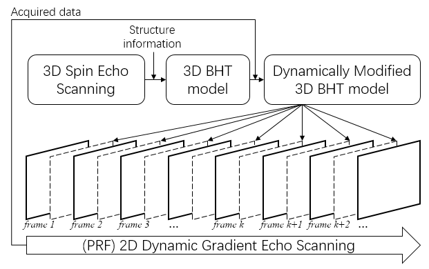
MR scanner (Philips Achieva, Philips Healthcare). As Fig.1 illustrates, T1 weighted 3D spin echo sequence (TE = 10ms, TR = 600ms, flip angle = 70$$$^{\circ}$$$) was firstly
used to acquire the 3D structure information of the phantom. Then a 2D gradient echo sequence (TE = 13ms,
TR = 30ms, flip angle = 20$$$^{\circ}$$$, FOV = 160mm$$$\times$$$160mm, resolution = 1mm) was used to acquire the temperature map of the transverse profile with a temporal resolution of 3.5s. Meanwhile the BHT model was performed with the structure information and the parameter $$$\lambda$$$ and $$$\phi$$$ are dynamically updated.
Results and Discussion
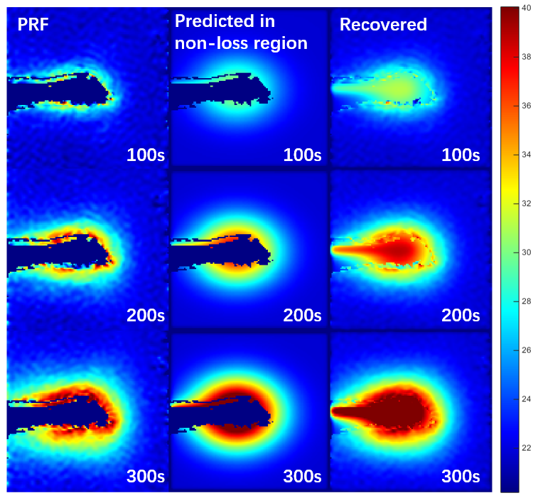
Fig.2 shows the PRF acquired map (column 1), predicted map in non-signal loss region (column 2) and recovered map (column 3) on 100, 200 and 300 seconds. PRF temperature imaging suffers much signal loss in the region of highest thermal dose, while the proposed method recovers the signal loss.
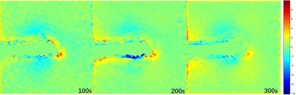
Fig.3 presents the error map between PRF acquired temperature map and predicted map in non-signal loss region on 100, 200 and 300 seconds. The RMSE between PRF acquired map and BHT predicted map in non-signal loss region of 100, 200 and 300 seconds is 1.54°C, 1.84°C and 1.32°C.
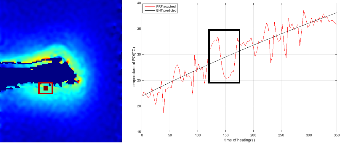
To demonstrate the improved accuracy of the proposed
method, a point of interested (POI) inside the red square in Fig.4 is erased and simulated as the signal loss point, the proposed method is performed to recover the temperature of the POI, then the temperature changing curve is plotted and compared to the temperature changing curve that acquired from PRF mapping. It is illustrated in black square that traditional PRF method is affected much by random errors due to noise, which leads to variation up to 8$$$^{\circ}$$$C, while the proposed method can recover a more accurate and smooth temperature changing.
Conclusion
A dynamically modified BHT enhanced PRF method was proposed. Phantom experiment shows that the proposed method can recover the signal loss caused by microwave probe. Temporal resolution could be increased to 1s or 0.5s with proper computing capacity.Acknowledgements
This work was supported by NSFC F012501.References
1. Ishihara Y, Calderon A, Watanabe H, Okamoto K, Suzuki Y, Kuroda K, et al. A precise and fast temperature mapping using water proton chemical shift. Magn Reson Med. 1995 Dec; 34(6):814–823.
2. Viola Rieke, Ron Instrella, Jarret Rosenberg, William Grissom, et al. Comparison of temperature processing methods for monitoring focused ultrasound ablation in the brain. J Magn Reson Imaging. 2013 Dec; 38(6).
3. Nick Todd, Allison Payne, Dennis L. Parker. Model predictive filtering for improved temporal resolution in MRI temperature imaging. Magn Reson Med. 2010 Apr; 1269-1279.
Figures