5427
MR-HIFU mild hyperthermia for recurrent rectal cancer: updated results from a phase I clinical trial1Radiation Oncology, Sunnybrook Health Sciences Centre, University of Toronto, Toronto, ON, Canada, 2Clinical Sites Research Program, Philips Research, Cambridge, MA, United States, 3Thunder Bay Regional Research Institute, Thunder Bay, ON, Canada, 4Electrical Engineering, Lakehead University, Thunder Bay, ON, Canada, 5Physical Sciences Platform, Sunnybrook Research Institute, Toronto, ON, Canada, 6Radiation Oncology, Sunnybrook Health Sciences Centre, Toronto, ON, Canada
Synopsis
We present the updated results of our phase I trial that delivers mild hyperthermia using magnetic resonance-guided high intensity focused ultrasound (MR-HIFU) combined with radiation and chemotherapy in the treatment of locally recurrent rectal cancer. Participation in the study is based on careful consideration of patient and tumor factors. MR-HIFU mild hyperthermia was delivered in three sessions during a 17-day regimen. MR-HIFU mild hyperthermia was successfully delivered and the procedure was well tolerated. No adverse effects have been reported following the combined treatment to date.
Introduction
Patients with locally recurrent rectal cancer who are ineligible for salvage surgery have limited outcomes when treated with re-irradiation and chemotherapy1. Hyperthermia may improve local control and quality of life in patients with local recurrence because of its potential to sensitize tumors to radiation and chemotherapy2. Here we report our updated clinical experience utilizing MR-HIFU to deliver mild hyperthermia treatment as an adjuvant to radiotherapy and chemotherapy in the context of locally recurrent rectal cancer.Methods
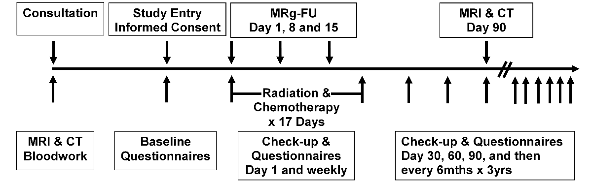
The Research Ethics Board of Sunnybrook Health Sciences Centre approved this clinical study. The standard of care for locally recurrent rectal cancer at our centre is the delivery of fractionated radiotherapy over 17 days (1.8 Gy/day) for a total of 30.6 Gy with concurrent fluoropyrimidine-based chemotherapy. In our study, three sessions of MR-HIFU mild hyperthermia are added to the standard treatment protocol, and delivered on days 1, 8, and 15 immediately before radiotherapy. Each hyperthermia session consists of positioning the patient on the MR-HIFU system, acquiring T1 and T2-weighted treatment planning images, and performing mild hyperthermia sonications under MR thermometry control with a target temperature and treatment time of 42°C for 30 minutes. Figure 1 outlines the study design.
MR-HIFU mild hyperthermia was delivered using the Sonalleve MR-HIFU system integrated into an Achieva 3T MRI (Philips Healthcare). Investigational Testing Authorization was granted by Health Canada to use a modified version of the treatment software adapted for large volume mild hyperthermia3,4. An electronically steered treatment cell with diameter of 18 mm was used to focus the ultrasound beam and cover the target tissue. Temperature maps used for feedback control were calculated dynamically using the proton resonance frequency shift technique with 3D first-order drift correction, in 6 image planes acquired using a water-selective T1-FFE-EPI sequence.
Results
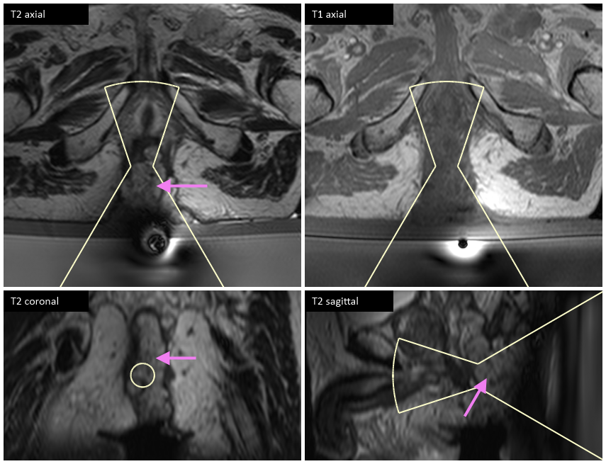
To date, two of eight screened patients have been enrolled. Six patients were not offered MR-HIFU by the study committee due to patient and tumor factors. The second patient had an inoperable T3 tumor with a large exophytic component extruding out of the anus measuring at least 5 x 8 x 4 cm. To achieve a favorable acoustic path covering a large portion of the tumor, the patient was positioned in an inclined supine orientation. Acoustic contact was achieved using stacked ultrasound gel pads and careful application of liquid ultrasound gel directly onto the tumor. Figure 2 depicts the planned treatment area on T1 and T2-weighted planning images.
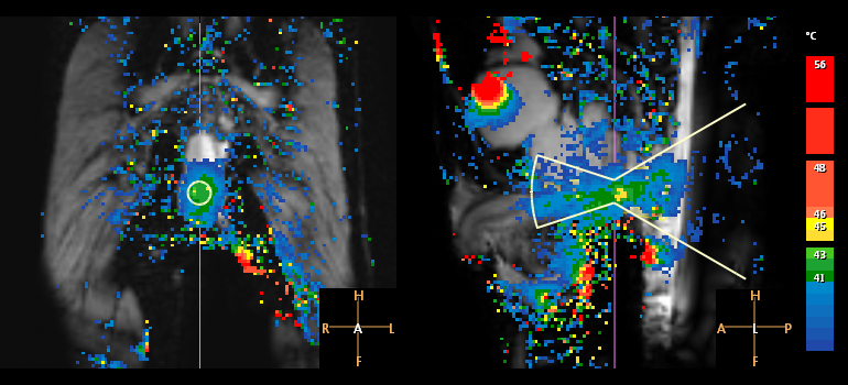
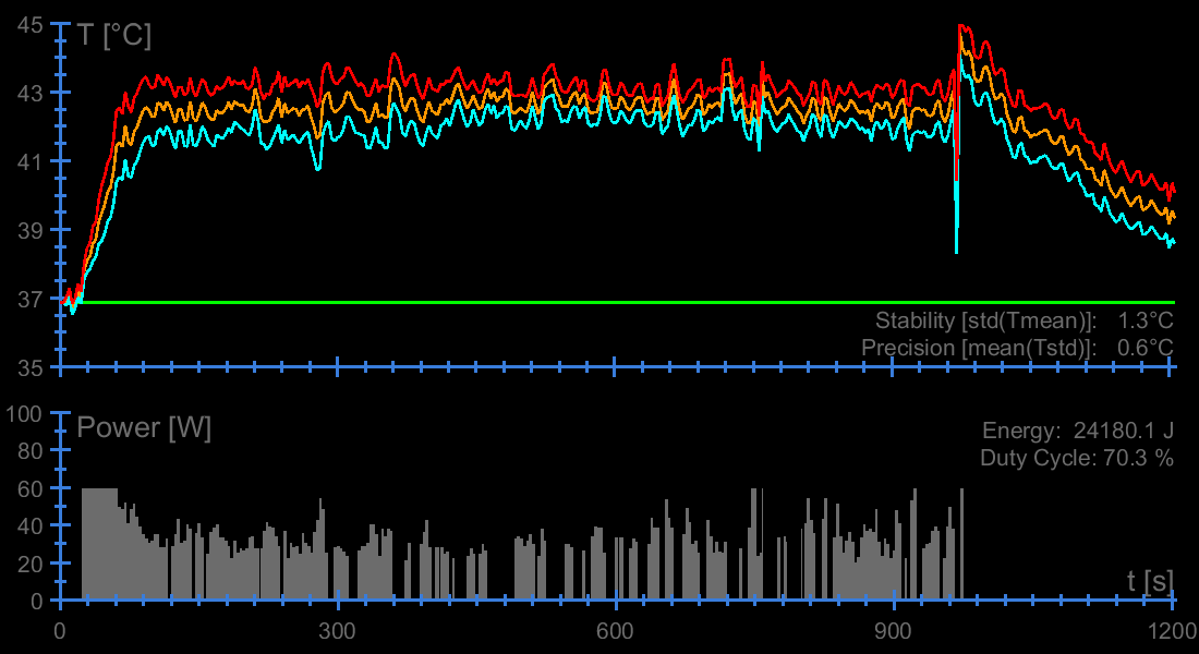
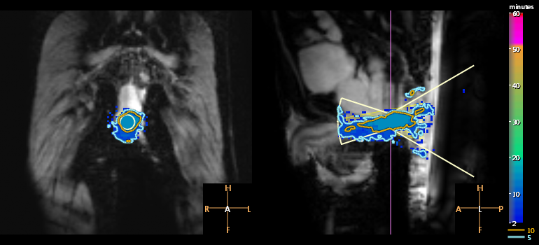
In his first HIFU session, an overall heating duration (time that target temperature was in the range of 40 to 45°C) of 31 minutes was achieved, using three hyperthermia sonications at 40-60 W and 1.0-1.2 MHz. Temperature mapping precision, evaluated as the spatial average of the temporal standard deviation in unheated pixels outside of the target area, was 1.0 ± 0.4°C across the three sonications with duration longer than 3 minutes. The longest sonication achieved average temperatures of 42.4°C across the 18 mm target region over 15 minutes, with temperatures exceeded by 90% and 10% of the target volume (T90 and T10) of 41.2 and 43.3°C suggesting uniform coverage (Figures 3 and 4). The volume heated to between 40 and 45°C for a therapeutic duration of longer than 10 minutes had a width and length of 27 and 71 mm (Figure 5). Post-treatment T2-weighted images did not indicate any unintended tissue damage.
While treatment was delivered, the patient was given light sedation, and he did not report any sensations of heating, nerve stimulation, or pain. The patient did not experience any other acute adverse events, and received his radiotherapy fraction within one hour of hyperthermia.
Discussion
Average temperatures of 42°C achieved in this second patient were higher than the best mean target region temperatures in a first patient (40-41°C)5. These differences are attributable primarily to differences in target location, as the same acoustic power and frequency were used. While the first patient had a deep seated tumor with skin, fat, and muscle between the transducer and the target, the patient reported here had a tumor which extended out of the body so that ultrasound propagated directly into the tumor.Conclusion
This update further demonstrates the technical and clinical feasibility of delivering mild hyperthermia using MR-HIFU under closed-loop MR-thermometry control.Acknowledgements
Financial support received from Canadian Cancer Society and Federal Economic Development Agency for Southern Ontario. Robert Staruch is a paid employee of Philips.References
1. Enríquez-Navascués JM, Borda N, Lizerazu A, et al. Patterns of local recurrence in rectal cancer after a multidisciplinary approach. World J Gastroenterol. 2011;17(13):1674-1684.
2. Kakehi M, Ueda K, Mukojima T, et al. Multi-institutional clinical studies on hyperthermia combined with radiotherapy or chemotherapy in advanced cancer of deep-seated organs. Int J Hyperthermia. 1990;6(4):719-740.
3. Tillander M, Hokland S, Koskela J, et al. High intensity focused ultrasound induced in vivo large volume hyperthermia under 3D MRI temperature control. Int J Hyperthermia. 2016;43(3):1539-1549.
4. Chu W, Staruch RM, Pichardo S, et al. Magnetic resonance-guided high-intensity focused ultrasound hyperthermia for recurrent rectal cancer: MR thermometry evaluation and preclinical validation. Int J Radiation Oncol Biol Phys. 2016;95(4):1259-1267.
5. Chu W, Staruch RM, Pichardo S, et al. MR-HIFU mild hyperthermia for sensitization of radiation and chemotherapy for recurrent rectal cancer: First phase I clinical trial results. ISMRM 2016;0824.
Figures