5399
Investigation of the relationship between metabolic basis of thalamus and brain spontaneous activity revealed by a study combined fMRI and MRS1Radiology, Nanjing Drum Tower Hospital, The Affiliated Hospital of Nanjing University Medical School, Nanjing, People's Republic of China
Synopsis
Several recent studies have reported a correlation between regional glutamate concentration and BOLD activation. In this study, we combined resting-state fMRI and MRS to investigate whether this association maintain in spontaneous brain activity in the thalamus. Significant positive correlation was found between glutamate concentration and the ALFF and ReHo in left thalamus; negative correlation between glutamate and DC in left thalamus. Furthermore, the ALFF of left primary motor cortex and bilateral auditory cortex were affected by left thalamus glutamate. This provides insight into better understanding the neuronal and biochemical mechanisms of thalamus function.
INTRODUCTION
The functions of thalamus are the relaying of sensory and motor signals to the cortex, and the regulation of consciousness(1). Recent studies combining biochemical and functional imaging measures have indicated that baseline glutamate and γ-aminobutyric acid (GABA) concentrations are associated with stimulus induced BOLD signal changes in regions such as visual cortex, and motor cortex(2). Fluctuations in spontaneous neural activity during rest consume most of the brain’s energy (3). Given that the majority of brain energy consumption is dedicated to support glutamatergic signaling (4), and so one would expect that the most energy intensive system - the glutamatergic- would be most related to resting state BOLD signal. Studies have demonstrated that glutamate levels in the posteromedial cortex were positively correlated with the intrinsic functional connectivity of the default mode network (5). Until now, the links between the local resting-state brain activity (ALFF, ReHo) and glutamate concentration are still largely unknown. In this study, we combined resting-state fMRI and MRS to investigate whether this association maintain in spontaneous brain activity in the thalamus.METHODS
We performed short echo Magnetic Resonance Spectroscopy (MRS) measurements of glutamate and resting-state fMRI in 17 young adult healthy participants. Bilateral thalami were selected as ROIs for MRS. The glutamate concentration of the thalamus was analyzed by Lc Model. The whole brain amplitude of low-frequency fluctuation (ALFF), regional homogeneity (ReHo) and degree centrality (DC) were calculated. The glutamate concentrations of bilateral thalamus were correlated with the whole brain ALFF, ReHo and DC.RESULTS
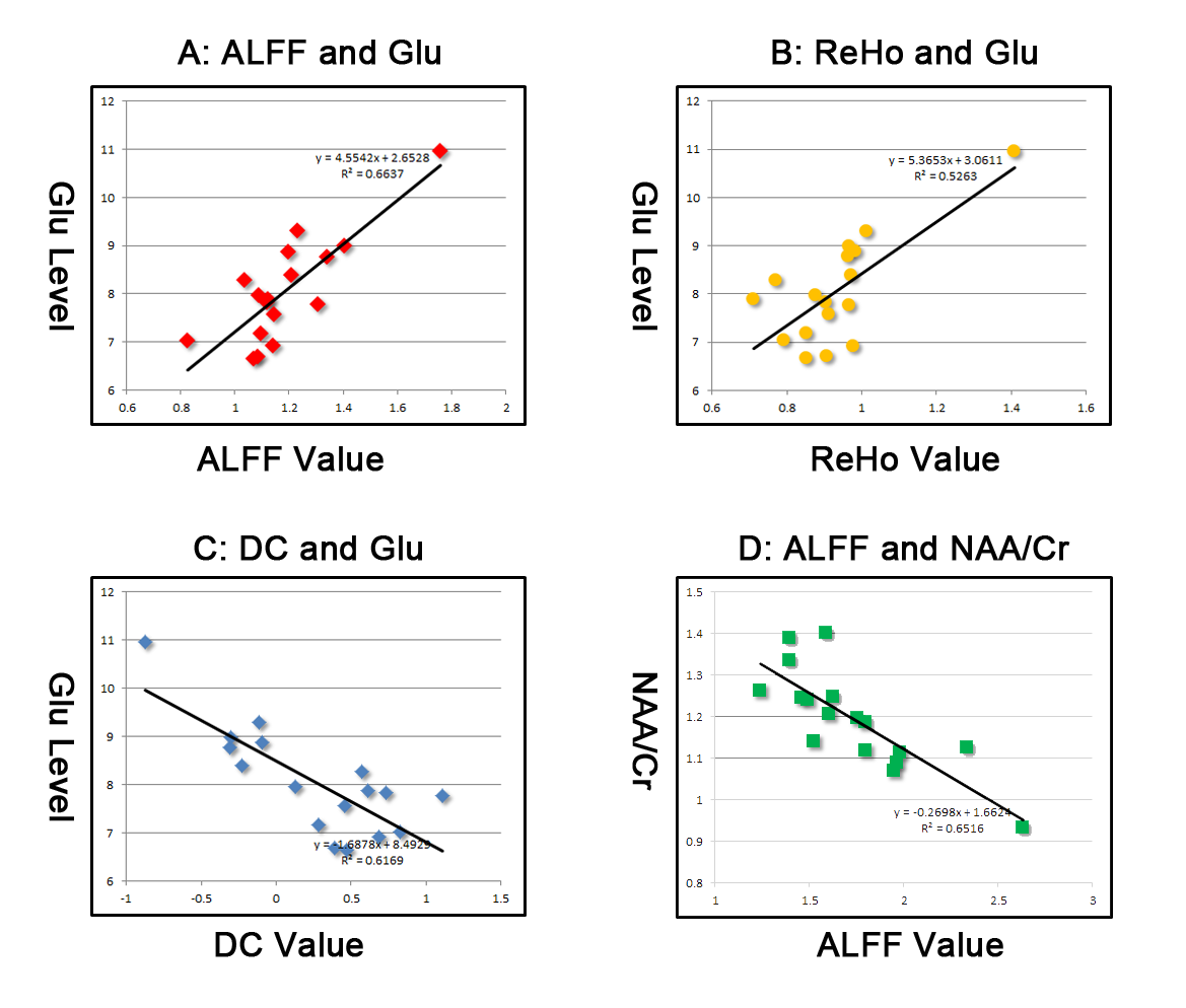
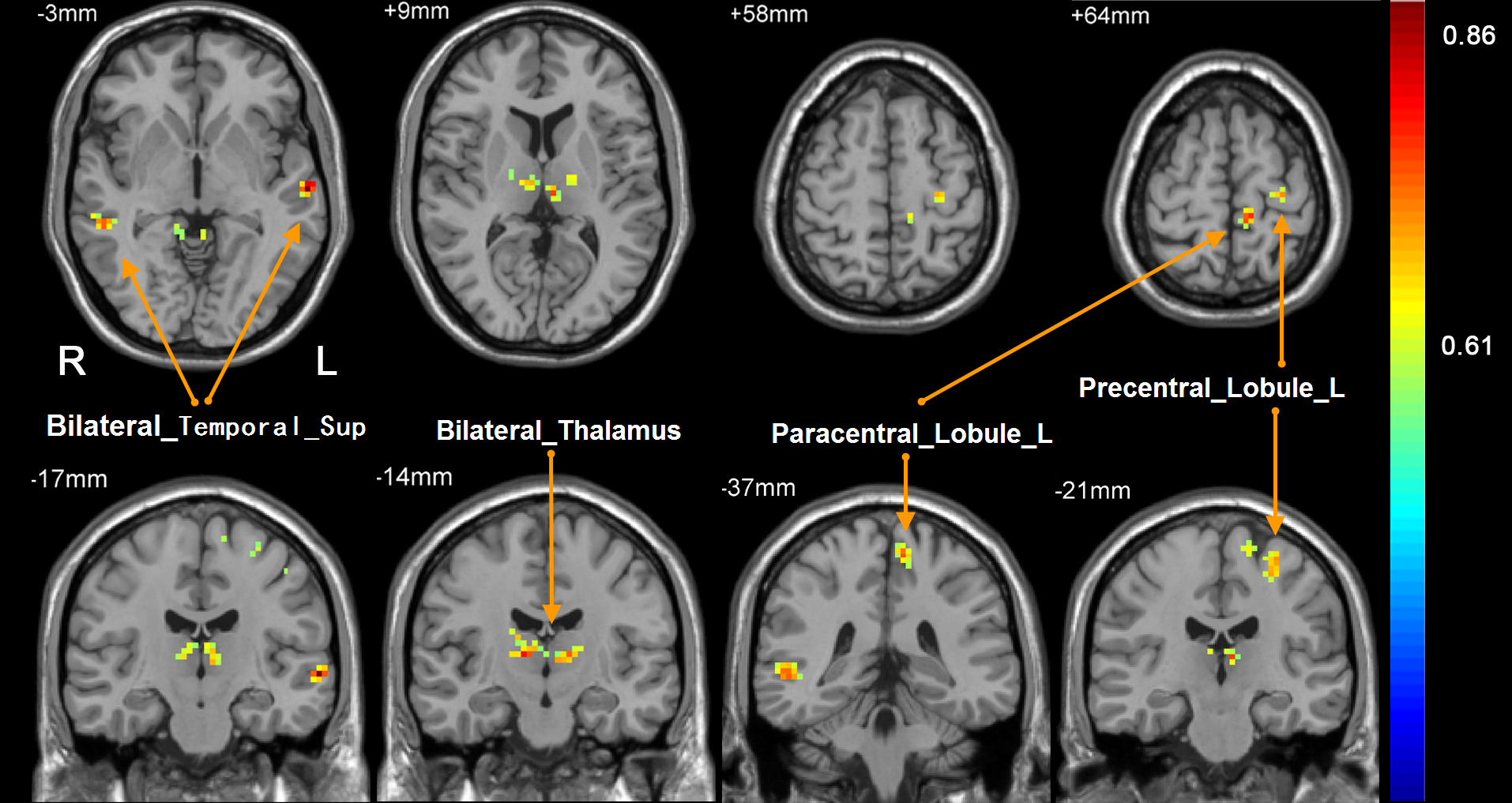
Significant positive correlation was found between glutamate concentration and the ALFF and ReHo in left thalamus; negative correlation between glutamate and DC in left thalamus. Furthermore, the ALFF of left primary motor cortex and bilateral auditory cortex were affected by left thalamus glutamate. However, left thalamus glutamate has an effect on the ReHo of left limbic system. No correlation was detected in right thalamus.DISCUSSION
Neural activity in general is constituted to a large degree by the excitation-inhibition balance. The balance impacts local hemodynamics, and is reflected in the BOLD signal (6). Our results showed positive correlation between glutamate concentration and ALFF level during resting state in the thalamus. ALFF measures the deviation of the BOLD signal, and reflects brain activity level during a period of time (7). And regional glutamate release can increase the metabolic rate and lead to regional BOLD signal increase. So, the results of our study can be interpreted as that glutamate regulates resting state ALFF level. We also found positive correlation between glutamate concentration and ReHo level during resting state. Another main finding of the current work is that glutamate levels in left thalamus are positively correlated with ALFF in left precentral gyrus, left postcentral gyrus and left paracentral lobule. It is well known that thalamus is a core structure of the brain and has specific relay nuclei that connect with distinct zones of the cortex (8). So we speculate that there are two reasons for regulation of ALFF levels in left precentral and postcentral gyrus by left thalamus glutamate: 1). They have structure connectivity. 2) Projections between them are glutamatergic.CONCLUSION
These results demonstrated that the excitability of thalamus decided its resting-state brain activity and has influence on the ALFF of sensory and motor cortex and the ReHo of limbic system. This provides insight into better understanding the neuronal and biochemical mechanisms of thalamus function under normal conditions and neuropsychiatric disorders.Acknowledgements
This study was supported by National Natural Science Foundation of China (Grant nos. 81301198, 81471643, 81571040, 81300925, 81422022, 81271553), the Natural Science Foundation of Jiangsu Province (BK20131085, BZ), the Provincial postdoctoral project (1501076A, BZ), the project of the sixth peak of talented people (WSN-O50, BZ), the key project of Nanjing Health Bureau (ZKX14027, BZ). We thank the volunteers for participating in this study. We thank the anonymous reviewers for their constructive suggestions to improve this work.References
1. Sherman SM. Thalamus plays a central role in ongoing cortical functioning. Nat Neurosci 2016;16(4):533-541.
2. Duncan NW, Wiebking C, Northoff G. Associations of regional GABA and glutamate with intrinsic and extrinsic neural activity in humans-A review of multimodal imaging studies. Neurosci Biobehav Rev 2014;47C:36-52.
3. Fox MD, Raichle ME. Spontaneous fluctuations in brain activity observed with functional magnetic resonance imaging. Nat Rev Neurosci 2007;8(9):700-711.
4. Hyder F, Patel AB, Gjedde A, Rothman DL, Behar KL, Shulman RG. Neuronal-glial glucose oxidation and glutamatergic-GABAergic function. J Cereb Blood Flow Metab 2006;26(7):865-877.
5. Kapogiannis D, Reiter DA, Willette AA, Mattson MP. Posteromedial cortex glutamate and GABA predict intrinsic functional connectivity of the default mode network. Neuroimage 2013;64:112-119.
6. Logothetis NK, Pauls J, Augath M, Trinath T, Oeltermann A. Neurophysiological investigation of the basis of the fMRI signal. Nature 2001;412(6843):150-157.
7. Zang YF, He Y, Zhu CZ, et al. Altered baseline brain activity in children with ADHD revealed by resting-state functional MRI. Brain Dev 2007;29(2):83-91.
8. Zhang D, Snyder AZ, Fox MD, Sansbury MW, Shimony JS, Raichle ME. Intrinsic functional relations between human cerebral cortex and thalamus. Journal of Neurophysiology 2008;100(4):1740-1748.
Figures