5397
Empirical Model Decomposition Removes Non-stationary EEG Noise in Simultaneous fMRI-EEG Acquisition1Aim for the Top University Project Office, National Taiwan Normal University, Taipei, Taiwan, 2Institute of Neuroscience, National Yang-Ming University, Taipei, Taiwan, 3Institute of Biomedical Engineering, National Taiwan University, Taipei, Taiwan, 4Department of Neuroscience and Biomedical Engineering, Aalto University, Espoo, Finland
Synopsis
Non-stationary EEG noise from simultaneous fMRI-EEG acquisition could be conventionally removed by optimal basis selection and followed by a low-pass filtering. An empirical model decomposition (EMD) method was applied to partially remove non-stationary EEG noise from simultaneous fMRI-EEG acquisition. Our results suggested that EMD method could reveal similar auditory evoked potential with optimal basis selection and low-pass filtering without prior knowledge or cut-off frequency, thus preserving high frequency signal not empirically related to the non-stationary noise. This EMD method allows us to investigate human brain high frequency EEG oscillation in the simultaneous fMRI-EEG measurement.
INTRODUCTION
Simultaneous EEG and fMRI acquisition provides a unique opportunity to investigate the neuronal basis of hemodynamic response in human brain (for review, see 1) with respectively high temporal and spatial resolution. However, this simultaneous acquisition could contaminate the EEG signal with the stationary noise, such as gradient artifact and pulse artifact from the MR imaging, and the non-stationary noise, such as ballistocardiogram (BCG) artifact and head movement from subject. The stationary noise has been commonly addressed by subtracting an averaged artifact template2 from the contaminated EEG measurements. The non-stationary noise, on the other hand, has been commonly addressed by optimal basis selection (OBS)3 and followed by a zero-phase low-pass filtering (LPF). One needs to manually select the numbers of optimal basis for OBS and the cut-off frequency for the low-pass filter, and also needs to know the R-R interval of the ECG when applied OBS. These aforementioned manual selections and prior knowledge can limit the performance of OBS, and the applications to the higher frequency oscillation in human brain.
Here we explore the possibility to remove the non-stationary EEG noise in the simultaneous fMRI-EEG acquisition without any prior knowledge or manual selections. Specifically, the empirical model decomposition (EMD)4 decomposed contaminated EEG signal into a set of intrinsic mode functions (IMF), and the clear EEG signal was composed again by a blindly selecting IMFs according to the themselves frequency spectrums. Experimental results show that our artifact removal strategy revealed cleaner auditory event-related potential (AEP) than OBS method. Our artifact removal strategy allows for ERP similar to that of OBS followed by LPF without a specific cut-off frequency.
METHODS
Volumetric EPI in a 3T scanner (Tim Trio, Siemens Medical Solutions, Erlangen, Germany) with TR of 2 sec was sampled concurrently with EEG signal. EEG (31 electrodes with impedance < 10 kΩ, reference electrode = FCz) was recorded continuously by a MRI-compatible system (BrainAmp MR Plus, Brain Products GmbH) with 5 kHz sampling rate. EEG was temporally synchronized to EPI via a 10 MHz clock in the MR scanner. Auditory stimulus (440 Hz; 200 ms duration) was delivered randomly between 0.5 and 1.5 s after the onset of each TR. There were 75 trials randomly distributed over a 5-minute scan. EPI were pre-processed by a customized stream (https://git.becs.aalto.fi/bml/bramila), and further analyzed by General Linear Model using canonical models to estimate hemodynamic response. The significance of BOLD signals (t-statistics) was morphed to inflated hemispheres of a standard template (fsaverage in FreeSurfer).
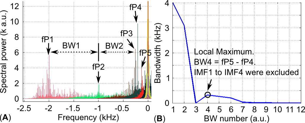
The EEG analysis started by first removing gradient artifacts using a MRI artifact template estimated directly from EEG-EPI recording. Subsequently, EEG signals removed the global trend and re-reference to the common averaged reference. EMD was applied on the EEG signal in each TR of fMRI to extract IMFs. Fourier Transform transformed IMFs into the frequency domain, and the frequency of the maximum spectral power (fPs in Figure 1A) for each IMF frequency was estimated. IMFs were categorized into groups by finding the local maximum of bandwidths between two adjacent IMF peak frequencies (Figure 1B). Clean EEG signal was reconstructed by excluding the IMFs belongs to higher frequency group. Epochs of EEG were created by taking 0.4 s and 1.0 s before and after each onset of auditory stimuli, respectively. For compares, re-referenced EEG signal was processed by OBS and 50 Hz LPF, and epochs and following AEP were created accordingly.
RESULTS AND DISCUSSION
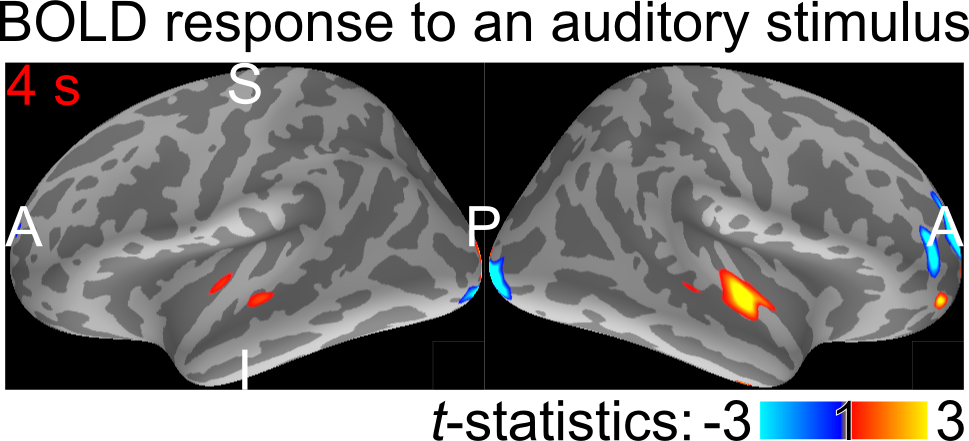
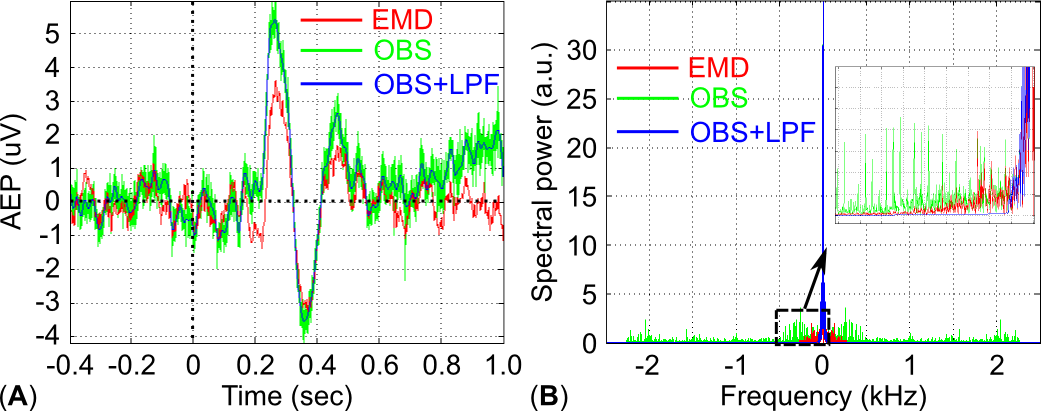
Figure 2 shows that significant hemodynamic activity was found at the bilateral superior temporal gyrus, suggesting that EPI normally detect the BOLD signal elicited by auditory stimuli. Figure 3A shows AEPs at the Cz electrode calculated by EMD, OBS, and OBS+LPF methods. These results suggest that AEPs with OBS only could still be noisy compared to that with EMD, while EMD and OBS+LPF on EEG-EPI measurements can generate similar and acceptable AEP. Importantly, fully data-driven EMD methods didn’t specify any cut-off frequency (Figure 3B) during the non-stationary noise removal so that the high frequency signal empirically suggested that not related to the non-stationary noise was still preserved.CONCLUSION
We proposed a fully data-driven method to estimate and remove the non-stationary EEG noise during the simultaneous fMRI-EEG acquisition without specify any prior knowledge or cut-off frequency. It is important to those experiments interested in cognitive functions, which are physiologically more related to neuronal oscillation beta (~ 20 Hz) and gamma (> 40 Hz) bands, since the frequency power spectrum of the non-stationary noise, e.g. BCG artifact, may alias up to 70 Hz. It is difficult to suppress the non-stationary noise by merely applying a LPF.Acknowledgements
The authors thank to Dr. Ching-Po Lin, and Dr. Li-Wei Ko for the hardware support.References
1. Ullsperger M. & Debener S. Simultaneous EEG and fMRI : recording, analysis, and application. (Oxford University Press, 2010).
2. Allen P.J., Josephs O., Turner R. A method for removing imaging artifact from continuous EEG recorded during functional MRI. NeuroImage 2000; 12:230-239.
3. Niazy R.K., Beckmann C.F., Iannetti G.D., et al. Removal of FMRI environment artifacts from EEG data using optimal basis sets. NeuroImage 2005; 28(3): 720-737.
4. Huang, N.E., Shen, Z., Long, S.R., et al. The empirical mode decomposition and the Hilbert spectrum for nonlinear and non-stationary time series analysis. Proceedings of the Royal Society of London A 1998; 454: 903-995.
Figures