5391
Diffusion kurtosis imaging with tract-based spatial statistics reveals white matter alterations in children with obstructive sleep apnea syndrome1Beijing Children's Hospital,Capital Medical University, Beijing, People's Republic of China, 2Chinese Academy of Sciences, Beijing, People's Republic of China, 3GE Healthcare, Beijing, People's Republic of China
Synopsis
It is reported that OSAS may cause cognitive function disorder of children due to chronic hypoxia for a long time[1]. However, it is not clear whether there are any structural changes of cerebral regions, e.g. white matter (WM). Diffusion kurtosis imaging is now widely used in the detection of cortical structural changes across kinds of diseases. In the present study, DKI is used to investigate microstructural changes of WM in OSAS children compared with normal healthy controls.
Introduction
Children with obstructive sleep apnea syndrome(OSAS)is the most common sleep-related breathing disorder is characterized by repeated episodes of partial (hypopnea) or complete (apnea) obstruction of the upper airway during sleep . It is reported that OSAS may cause cognitive function disorder of children due to chronic hypoxia for a long time[1]. However, it is not clear whether there are any structural changes of cerebral regions, e.g. white matter (WM). Diffusion kurtosis imaging is now widely used in the detection of cortical structural changes across kinds of diseases. In the present study, DKI is used to investigate microstructural changes of WM in OSAS children compared with normal healthy controls.Purpose
To understand the developmental cause of such changes,we investigated microstructural changes of WM in OSAS children by using Diffusion kurtosis imaging(DKI).Methods
Ten children diagnosed with OSAS were recruited in the present study (age: 5.4±2.221 years, range: 4–10 years; 6 female). For comparison, eleven age and gender matched healthy controls were also included MR images were acquired in Beijing Children’s Hospital using a 3.0 T GE Discovery MR750 scanner.For each subject, T1 weighted imaging (T1 WI),T2 weighted imaging (T2 WI) and DKI were scanned. And on conventional MRI,the imaging apearances of all the cases were normal.For DKI dataset, conventional kurtosis related parameters such as: Axial diffusivity (AD), Radial diffusivity (RD), Mean diffusivity (MD), Fractional anisotropy (FA), Kurtosis fractional anisotropy (KFA), Axial kurtosis (Kax), Radial kurtosis (Krad), Mean kurtosis (Kmean), Mean kurtosis tensor (MKT) were firstly computed for each subject. Then, Tract-Based Spatial Statistics (TBSS) was used to assess differences in WM between children with OSAS and normal controls.Results
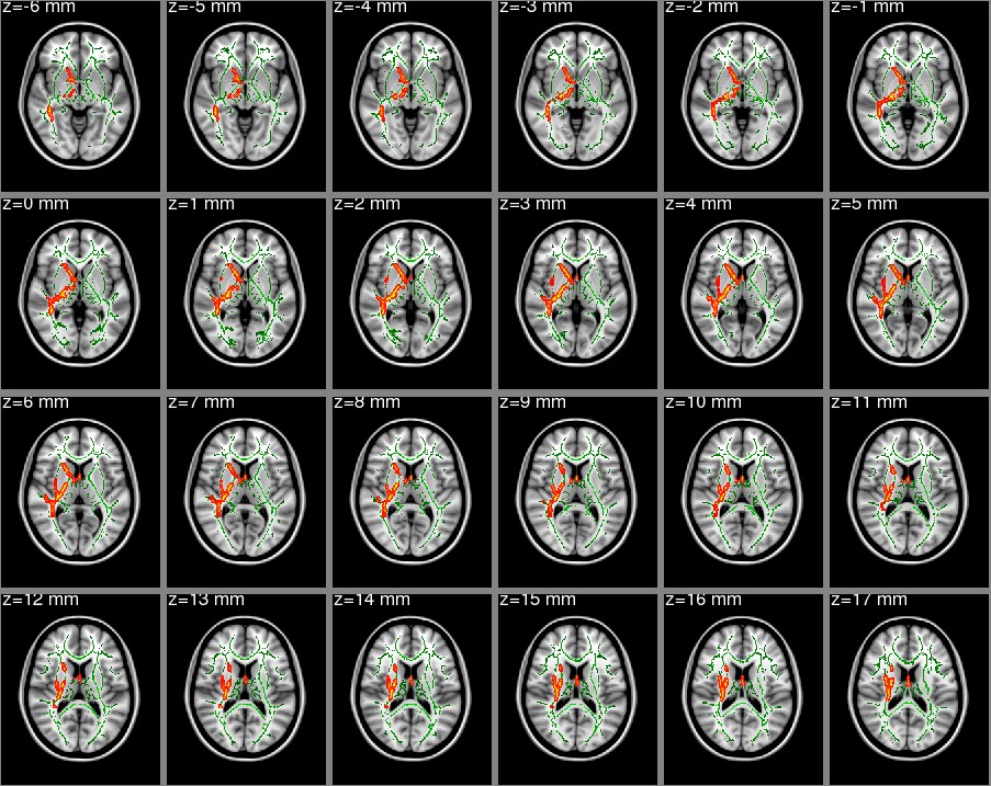
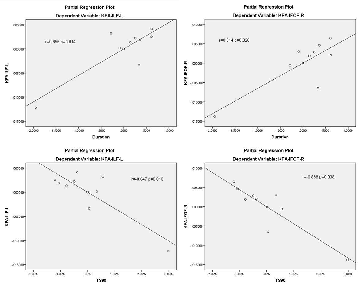
The white matter fibers with significant intergroup differences (FEW corrected P < 0.05) in DKI-derived kurtosis parameters are shown as bellows. Compared to healthy controls, the kurtosis parameters were exhibited significantly decreased in multiple white matter regions in OSAS children. The mainly involved region include bilateral corticospinal tract, inferior fronto-occipital fasciculus, inferior longitudinal fasciculus, posterior thalamic radiation (include optic radiation); right superior longitudinal fasciculus; left inferior frontal gyrus, brainstem, body of corpus callosum/forceps minor (Fig1,Fig2).In TBSS analysis of DKI parameters, KFA values in the left Inferior longitudinal fasciculus, right Inferior fronto-occipital fasciculus were significantly positively correlated with duration time of the patient group,and negativelycorrelated withTS90 (the percentage of the total recorded time when the oxygen staturation below 90%) of the patient group (Figure3).Discussion
Chronic repeated reversible low oxygen caused by OSAS will impact on children's cognitive behavioral development and academic.The adult of symptoms such as burnout happen less often in children, nerve behavior problems may be the better reflect in children with OSAS which characterized by the ability of learning and memory impairment and abnormal behavior, inattention and words can not express clearly, etc[2]. Childhood is an important stage of brain development mature, in terms of neural development, serious long-term damage can also occur cognitive function defects.Long-term under the condition of harmful to the environment, the function of the nervous tissue growth can produce periodic damage.OSAS were sensitive to oxygen in infants can cause irreversible brain damage OSAS easily affect the nerve center, the main affected areas include the frontal lobe, subcortical area, the hippocampus and extensive white matter nerve centre connected, easy to cause the functional defect in pathological change. Invalid or insufficient sleep can cause anterior frontal cortex dysfunction, such as behavioral inhibition, emotion regulation and working memory to perform functions such as damage, increase the distraction and hyperactivity[3]. Hypoxia stress in the pathogenesis of abnormal changes of neurotransmitters in the brain is extremely complex, oxygen free radicals caused by lipid peroxidation and molecular oxygen lack of neurotransmitter synthesis decomposition obstacle is the main cause of abnormal brain neurotransmitter levels.Animal experimental results prove that intermittent hypoxemia, neurons can be induced cell death, damage for some special memories[4]. There are many very meaningful results in this study:Based on the traditional DTI,value of FA could not find a positive result, but the value of KFA has a positive result,we can say that the value of KFA in multiple areas significantly lower than normal children. .The quantitative analysis of brain changes using DKI suggests the abnormalities of putamen and caudate nucleus in OSAS children patients,which may indicate the pathological change in OSAS children patients.Conclusion
DKI is more suitable than DTI for grasp tissue microstructure change, can better reflect the brain white matter changes in the microstructure.Children with OSAS showed significant functional alterations of the left Inferior longitudinal fasciculus, right Inferior fronto-occipital fasciculus and longitudinal fasciculus in children with OSAS.Acknowledgements
No acknowledgement found.References
[1] HalbowerAC;DeganokarM;BarkerPB.Childhood obstructive sleep apnea associates with neuropsychological deficits and neuronal brain injury[J].Pies Med,2006,3:e301.
[2] Zhang,Y,Ma,Q,Liu,XT.Study on sleep and respiratory characteristics and effect on neurocognitive in children with obstructive sleep apnea syndrome in different age[J].Journal of Clinical and Experimental Medicine,2015,(15):1301-1303.
[3] Farre R,Montserrat J.Navajas D.Merbidity due to obstructive sleep apnea:insights from animal models.CurrOpinPulm Med,2008,14(6):530-536.
[4]Zhang J,Fung S,Xi M,et ai.Recurrent apnea induces neuronal apoptosis in the guinea pig fnrebrain.ExpNeuroi,2009,2 1 6(2):290.294.
Figures
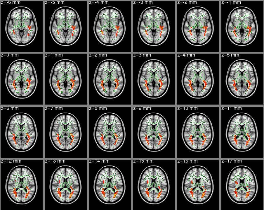
Kurtosis fractional anisotropy (KFA), degree of anisotropy of the kurtosis tensor Controls > patients (cluster size >20):Figure 1. Skeleton clusters showing significantly decreased Kfa at p<0.05