5386
White Matter Resting-State fMRI with Hypercapnic Respiratory ChallengeTung-Lin Wu1,2, Jennifer Watchmaker1, Li Min Chen1,3, Adam W Anderson1,2,3, Zhaohua Ding1,2,3, and John C Gore1,2,3
1Vanderbilt University Institute of Imaging Science, Nashville, TN, United States, 2Biomedical Engineering, Vanderbilt University, Nashville, TN, United States, 3Radiology and Radiological Sciences, Vanderbilt University, Nashville, TN, United States
Synopsis
In order to further elucidate the biophysical origins of spatio-temporal correlation tensors and validate the possibility of detecting BOLD signals in white matter, we acquired resting-state fMRI in volunteers breathing alternately room air and CO2 enriched air to induce a hypercapnic-normoxic change in CBF and CBV. Our hypercapnic respiratory challenge experiments suggest that spatio-temporal correlations in white matter may be driven by local hemodynamic effects, consistent with BOLD effects instead of other potential mechanisms. Our results also imply and support our previous observation that BOLD signals in white matter can be reliably detected, and resting-state correlations between voxels are anisotropic.
Target Audience
Researchers interested in studying brain function, white matter and fMRI.Background
Over the past two decades, numerous studies have used resting state functional magnetic resonance imaging (rsfMRI) to characterize functional connectivity between cortical regions. To date, however, there have been relatively few studies reporting blood oxygenation level dependent (BOLD) signals in white matter, and only a handful looked at resting state fluctuations. However, a number of investigations have demonstrated reliable BOLD signal detections in white matter, and we recently reported the detection of anisotropic resting state correlations within white matter that appear to depict an underlying structure.1 We introduced the concept of spatio-temporal correlation tensors that are able to delineate functional pathways in white matter purely on the basis of rsfMRI data2 and these functional structures are often similar to those obtained from DTI data.3 Moreover, we have found white matter BOLD signal fluctuations behave similarly to those in gray matter as baseline neural activity is altered with different anesthesia levels.4 In order to further elucidate the biophysical origins of spatio-temporal correlation tensors and validate the possibility of detecting BOLD signals in white matter, we acquired rsfMRI in healthy volunteers breathing alternately room air and CO2 enriched air to induce a hypercapnic-normoxic change in cerebral blood flow (CBF) and cerebral blood volume (CBV).Methods
MRI acquisitions were obtained using a 3.0 T Achieva scanner (Philips Healthcare) from 4 healthy volunteers. Participants wore a mask that covered the nose and mouth, and were imaged in a resting state with (i) room air administered for 6 minutes followed by (ii) hypercapnic-normoxia gas (5% CO2/21% O2/74% N2, HC-NO) for another 6 minutes per run. A total of 7 runs were acquired in this study with BOLD imaging performed using a single-shot gradient-echo EPI sequence (TE/TR=30/2000 ms, spatial resolution=3 x 3 x 3.5 mm3, SENSE-factor=2). Anatomical T1-weighted images were acquired using a 3D fast gradient echo sequence (TE/TR=4.6/8.9ms; spatial resolution = 1 x 1 x 1 mm3). fMRI pre-processing was performed using spm12, which included slice-timing correction, motion correction and isotropic smoothing of 3 x 3 x 3 mm3. Functional tensors were subsequently constructed from voxels based on calculations of the anisotropic resting state correlations with 26 nearest neighbors. The fractional anisotropy and major eigenvalues of the tensors at each voxel were computed. Details of the tensor construction method can be found in Ding et al.2 All studies were approved by the local Institutional Review Board.Results and Discussion
Representative averaged time series before and after the introduction of hypercapnic-normoxia gas are presented in Figure 1. Average signal increases of 2.55% and 1.16% were observed in gray and white matter respectively. The average MRI signal variance in the low frequency range (0.01-0.08Hz) increased by 11.66% and 13.94% in gray and white matter respectively after gases were administered. Averaged signal variance in white matter was also found to be approximately 45% of that in gray matter. Maps of spatio-temporal correlation tensors were subsequently constructed and compared before and after the change of breathing gases shown in Figure 2. Specifically, we observed tensor orientations in white matter became more consistent among neighboring voxels, and dominant directions of the tensors agreed better with local macroscopic tissue structure during hypercapnia, while tensors remained largely unaffected in gray matter. Figure 3 presents fractional anisotropy and major eigenvalues (i.e. greater nearest neighbor anisotropy of correlations) of the spatio-temporal correlation tensors in white matter from one representative subject. The absence of fractional anisotropy changes can be attributed to global increases in neighboring correlations, which is supported by the observation of greater eigenvalues and signal variance in the time series. Overall, these findings further confirm the presence of resting state BOLD signals in white matter, and that a change in CBF and CBV stimulates a change in their resting-state correlations. Importantly, our results here also suggest that spatio-temporal correlation patterns observed in white matter depend on local vascular effects as opposed to other possible confounding effects.Conclusion
Our hypercapnic respiratory challenge experiment suggests that spatio-temporal correlations in white matter may be driven by local hemodynamic effects, consistent with BOLD effects instead of other potential mechanisms. Our results also imply and support our previous observation that BOLD signals in white matter can be reliably detected, and resting state correlations between voxels are anisotropic. Finally, findings here may have further implications on assessing hemodynamic impairment in patients with cerebrovascular disease.Acknowledgements
No acknowledgement found.References
[1] Gawryluk, J.R. et al. (2014) Does functional MRI detect activation in white matter? A review of emerging evidence, issues, and future directions. Front. Neurosci. 8, 1–12. doi:10.3389/fnins.2014.00239 [2] Ding Z et al. (2013) Spatio-temporal correlation tensors reveal functional structure in human brain. PLoS One 8(12): e82107. [3] Ding Z et al. (2015) Visualizing functional pathways in the human brain using correlation tensors and magnetic resonance imaging. Magn. Reson. Imaging 34, 8–17. doi:10.1016/j.mri.2015.10.003 [4] Wu et al. (2016) Effects of anesthesia on resting state BOLD signals in white matter of non-human primates Magn. Reson. Imaging 34(9):1235-1241. doi: 10.1016/j.mri.2016.07.001.Figures
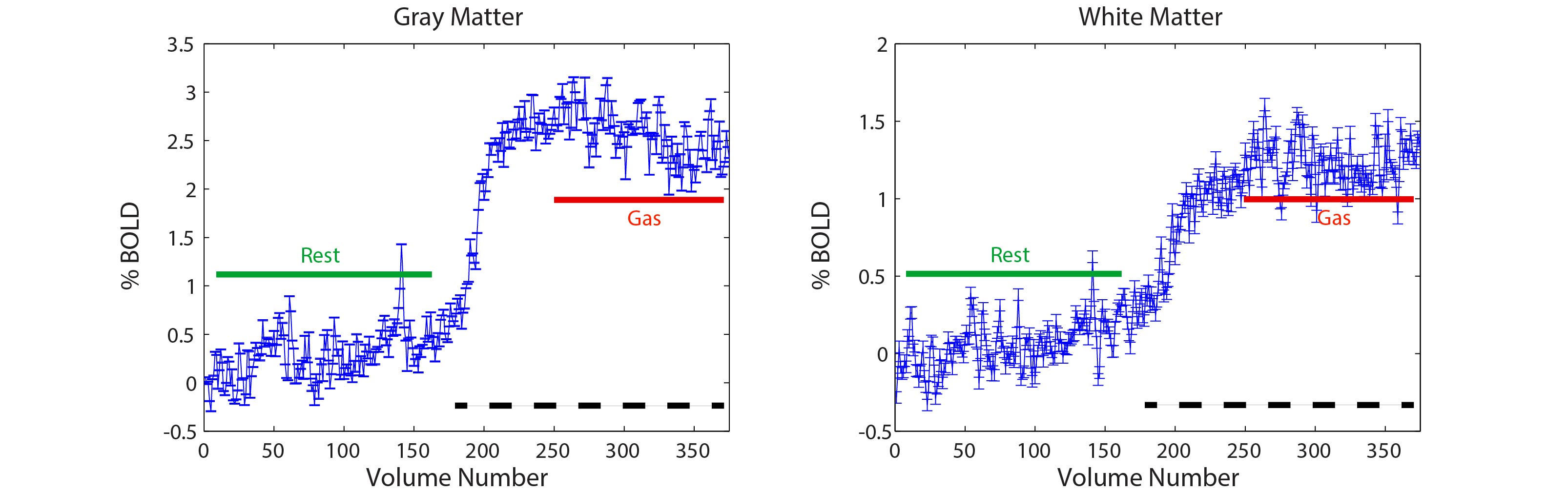
Representative averaged time series for gray and white matter. Green line represents
volumes extracted for ‘rest’, red line shows volumes extracted for ‘gas’ and
black dashed line indicates time period of gas administration. Averaged signal
fluctuations (variance) in the low frequency range (0.01-0.08Hz) increased by
11.66% and 13.94% in gray and white matter respectively after the introduction
of gases.
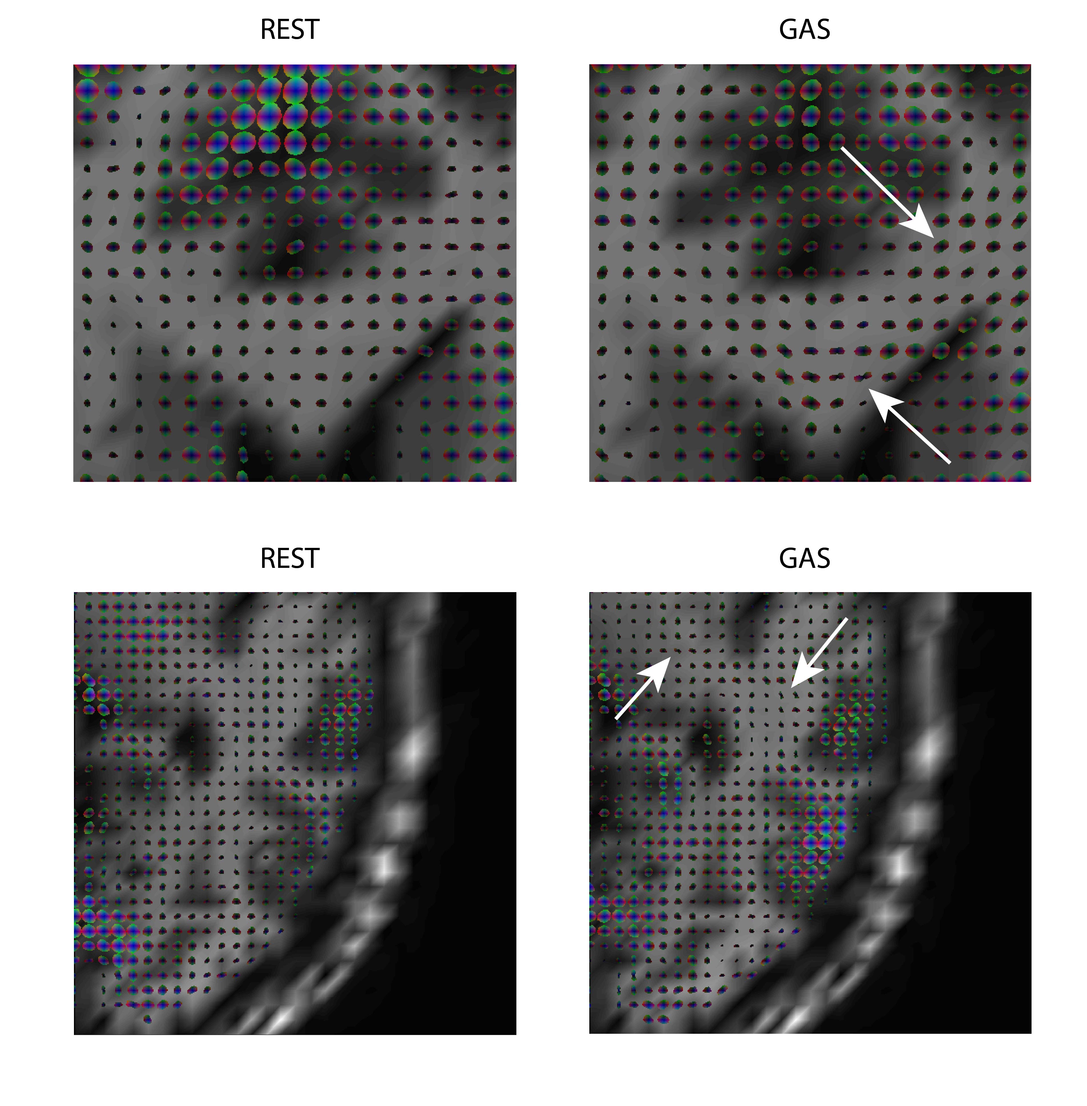
Spatio-temporal correlation tensor maps under ‘rest’
and ‘gas’ conditions from the same runs for comparison. With the introduction
of CO2, tensor orientations are observed to be more consistent among neighboring
voxels and dominant directions of the tensors agree better with local
macroscopic tissue structure (pointed out by the white arrows).
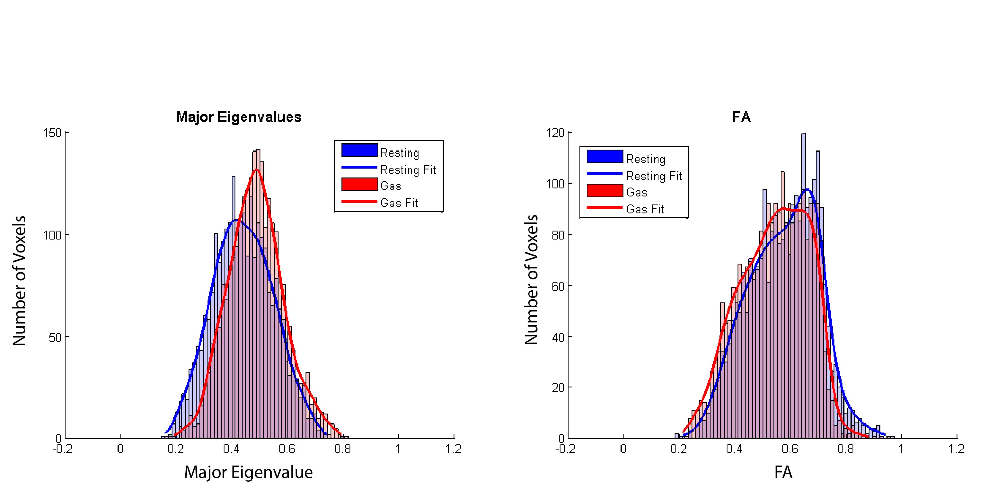
Histograms of
major eigenvalues and fractional anisotropy (FA) in white matter before (blue)
and after (red) introduction of CO2 gas in one representative
subject. An increase in major eigenvalue of the spatio-temporal correlation
tensor is observed while the FA remains relatively constant.