5376
The Early Global Function Connectivity Stability in Infants during the Neonatal Period1Department of Biomedical Engineering, Peking University, beijing, People's Republic of China, 2Center for MRI Research, Academy for Advanced Interdisciplinary Studies, Peking University, Beijing, People's Republic of China, 3Daping Hospital, Third Military Medical University, Daping Hospital, Third Military Medical University, Chongqing, People's Republic of China, 4MR Research China, GE Healthcare, Beijing, People's Republic of China, 5Biomedical Imaging Research Institute (BIRI), Department of Biomedical Sciences and Imaging, Cedars-Sinai Medical Center, los angeles, CA, United States, 6Department of Radiology and Biomedical Research Imaging Center, University of North Carolina Chapel Hill, United States
Synopsis
This study proposes an improved method named global functional connectivity stability (GFCS) to quantify the brain dynamic functional connectivity at a voxel-wise level. The GFCS was applied to investigate the overall functional connectivity stability and its correlation with time in infants during the period from late preterm to the term equivalent age (TEA). It is shown that infants presented high functional stability predominantly in the sensorimotor areas, temporal lobe, posterior cingulate cortex (PCC) and medial prefrontal cortex. With time, the frontal areas appeared more variable while the sensorimotor cortex appeared more stable in infants during the neonatal period.
Purpose
Functional brain networks demonstrated significant temporal variability. This temporal variability, although previously considered as noise1, now was increasingly recognized to be related to behavior, cognition and disease2, 3. Currently, most studies focused on the dynamic functional connectivity between a given pair of regions of interest4, or the connectivity of the whole brain5. In a recent study, Zhang and co-workers proposed a novel method to measure the overall functional connectivity variability of a given region6. However, the whole brain voxel-wise variability or stability of functional connectivity has not been investigated. Moreover, most work on brain dynamics is centralized in adults while the functional dynamic architecture of infants remains largely unexplored. In this study, a new index was proposed to assess voxel-wise stability of overall functional connectivity. In addition, this index was applied to investigate functional stability of infants’ brains during the period from the late preterm to term equivalent age (TEA).Methods
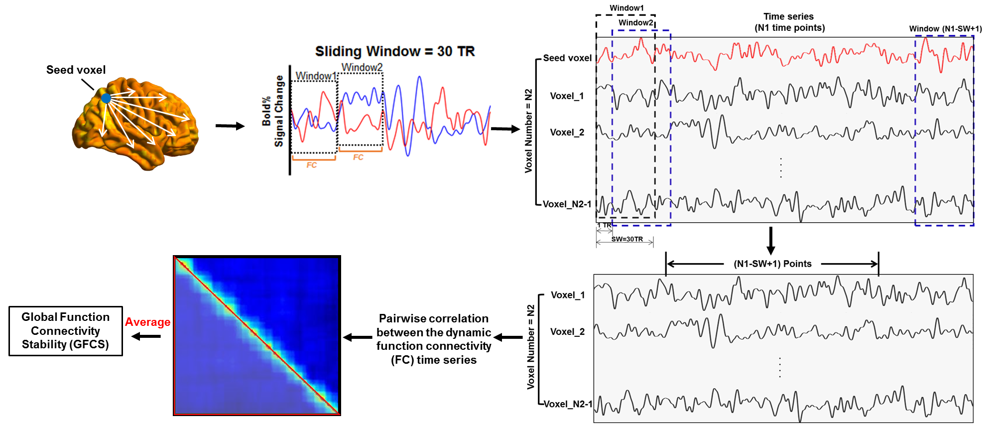
In this study, 22 normal preterm infants (gestational age, GA<37 wk) and 31 full-term infants (GA>37 wk) were included. An index named the global functional connectivity stability (GFCS) was proposed to assess the temporal stability of overall functional connectivity at a voxel-wise level. The computation of GFCS was shown in Fig 1. Specifically, for a given seed voxel, the windowed (window size is 30 volumes) correlation was calculated between the time series of this voxel and every other voxels of the whole brain. The window was iteratively moved forward with step size of one time point until the last 30 time points were selected. Hence, each seed had (N-30+1) windowed correlation maps (supposed the time series included N volumes). Within each window, the correlation map was reformed into a 1-D vector. Then, pairwise correlation was conducted amongst these 1-D vectors. Finally, a (N-30+1)*(N-30+1) similarity matrix characterizing the similarity between different windowed functional connectivity was obtained, and the averaged value of the similarity matrix was defined as the GFCS for a given seed voxel. Same procedures were extended to every other voxels, and the GFCS map of each subject was obtained (see Fig 1). To characterize the group-level stability pattern, averaged GFCS maps of the preterm, full-term infants and whole infants (both preterm and full-term) were calculated respectively. Furthermore, functional stability development was evaluated through calculating the voxel-wise correlations between GFCS maps and the postmenstrual ages (PMAs) of the whole infants. All statistical tests underwent AlphaSim correction (voxel-level p<0.05, cluster-level p<0.05). This mild multiple comparison correction was applied so as not to neglect the subtle changes that occurred at this critical period.Results and Discussion
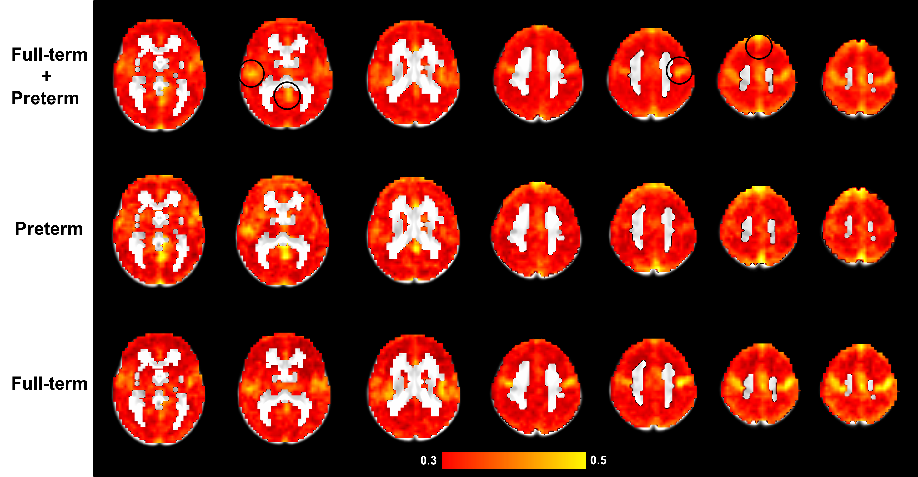
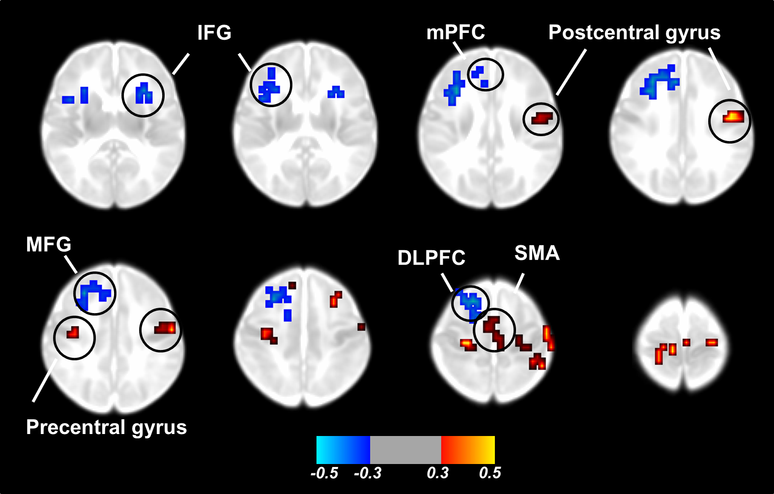
Mean GFCS maps of different groups of infants were shown in Fig 2. It can be seen that, GFCS map of whole infants showed high stability predominantly in the sensorimotor areas, temporal lobe, posterior cingulate cortex (PCC) and medial frontal cortex. Consistent results were reported in ref 66, where areas of sensorimotor cortex and default mode network (DMN) related regions appeared less variable over time. As indicated by their investigations, the temporal variability may reflect the flexibility and adaptability of brain function. Hence, areas with high stability in our results were probably indicative of a primary function (sensorimotor) or stable connectivity over time (DMN). The temporal lobe in their study demonstrated high variability, which was different from our results, possibly indicating that the functions of temporal areas were still primitive and would undergo further development. As compared to full-term infants, preterm infants exhibited decreased GFCS mainly in the sensorimotor cortex. Furthermore, with regard to the relationship between GFCS and PMAs, significant positive correlations were found in the sensorimotor cortex including precentral/postcentral gyrus, supplementary motor areas (SMA), while areas of inferior frontal gyrus (IFG), medial prefrontal cortex (mPFC), middle frontal gyrus (MFG), and dorsolateral prefrontal cortex (DLPFC) demonstrated significantly negative correlations (see Fig 3). These findings may reflect that the tendency for brain stability development in infants presents a heterogeneous topography in infants, i.e., the frontal areas appear more variable over time while unimodal areas, such as sensorimotor cortex, appear more stable during the neonatal period.Conclusion
Our results show that, infants present high functional stability predominantly in the sensorimotor areas, temporal lobe, PCC and medial prefrontal cortex. With time, frontal areas become more variable while sensorimotor cortex appears more stable in infants during the neonatal period. The GFCS may be a potentially useful measure to quantify the functional connectivity dynamics of the brain.Acknowledgements
This study was funded by the National Science Foundation of China (Grant No. 81173662 to Y.S.) and the Programs for the Development of Science and Technology of Chongqing in China (Grant No. 2011GGC083 to Y.S.). The authors have stated that they had no interests which might be perceived as posing a conflict or bias.References
1 Garrett, Douglas D., et al. Journal of Neuroscience the Official Journal of the Society for Neuroscience 30.14(2010):4914-21.
2 Calhoun, et al. Neuron 84.2(2014):262-74.
3 Kopell, et al. Neuron 83.6(2014):1319-28.
4 Chang, Catie, and G. H. Glover. Neuroimage 50.1(2010):81-98.
5 Allen, E. A., et al. Cerebral Cortex 24.3(2012):663-76.
6 Jie, Zhang, et al. Brain 139.8(2016).
Figures