5366
fMRI detection of neuromodulation induced neuroplasticity after spinal cord injuryVijai Krishnan1,2, Anna Schwartz1, William Stokes1, Jeff W.M. Bulte2, Jineta Banerjee2, Aline Thomas2, Pablo Celnik3, and Galit Pelled1,2
1F.M. Kirby Center, Kennedy Krieger Institute, Baltimore, MD, United States, 2Department of Radiology, Johns Hopkins University School of Medicine, Baltimore, MD, United States, 3Physical Medicine and Rehabilitation, Johns Hopkins University School of Medicine, MD, United States
Synopsis
Spinal cord injury (SCI) leads to severe motor and sensory deficits. New advances in non-invasive neuromodulation technologies such as transcranial magnetic stimulation (TMS) have shown promise in facilitating recovery following brain injuries. Here we tested whether TMS therapy can be developed as a rehabilitative approach in a rat model of SCI. High-resolution functional MRI (fMRI) at 11.7 T was used to detect cortical activity associated with post-injury neuroplasticity. A battery of behavioral tests was used to monitor gross changes in motor behavior. Our results demonstrate that TMS therapy is beneficial in improving post-SCI functional outcomes.
Introduction
Spinal cord injury (SCI) is the leading cause of disability causing partial or complete damage to sensory and motor pathways, thereby altering neural circuits. There are no rehabilitative strategies that have shown a robust improvement, and there are no imaging protocols to detect neuroplasticity associated with rehabilitation after spinal cord injury. Evidence from basic research suggests that within minutes after SCI, decreases in spontaneous neuronal activity are observed in cortical areas that correspond to the injured as well as to the non-injured limbs. Moreover, these decreases were correlated with poor recovery. Thus, we hypothesized that an intervention to attenuate the decreases in neuronal activity applied immediately post-SCI would accelerate neurorehabilitation. Transcranial magnetic stimulation (TMS) is a non-invasive method to induce neuronal excitation and plasticity beyond the stimulation period. We have recently demonstrated that following traumatic brain injury in rats TMS has rescued neuronal activity and led to improvement in behavioral tests (Lu et al., Scientific Reports, 2015). Therefore, we have determined if TMS can improve functional outcome post-SCI. Currently, the standard assessment of SCI in rats is limited to gross behavioral tests. These tests may not be sensitive to neuroplasticity changes associated with recovery. Here we used high-resolution fMRI obtained at 11.7 T to monitor if TMS therapy induces post-injury neuroplasticity. We determined the optimal TMS therapy protocol that leads to the greatest evoked- fMRI responses to limb stimulation. Our results demonstrate that fMRI may be a sensitive method to report on SCI neuroplasticity.Methods
SCI was induced at segment T7 in adult rats. This type of injury results in hind limb dysfunction. We tested sensorimotor function in three groups: SCI rats that started receiving TMS within 10 min after the procedure (immediate-TMS; n=7); SCI rats that received TMS starting two weeks after the procedure (delayed-TMS; n=5), and SCI rats that received sham TMS (no-TMS, n=7). High-frequency (20 Hz) TMS was applied to the sensorimotor cortex via a custom built rodent coil, and was delivered for 10 min, three times a week, for a total of six weeks. Sensory responses to 9 Hz hind limb stimulation were delivered to dexodormitor anesthetized rats. Blood-oxygenation-level-dependent (BOLD) fMRI responses to contralateral tactile limb stimulation were measured in an ultra-high field of an 11.7 T/16 cm horizontal bore small-animal scanner (Bruker BioSpin, Rheinstetten, Germany). Five 1 mm thick coronal slices covering S1 were acquired (effective echo with a field of view (FOV), 1.92 × 1.92 cm; matrix size, 128 × 128). A T2-weighted RARE sequence was used to acquire high-resolution anatomical images FOV, 1.92 × 1.92 cm; matrix size, 256 × 256) corresponding to the BOLD fMRI measurements.Results
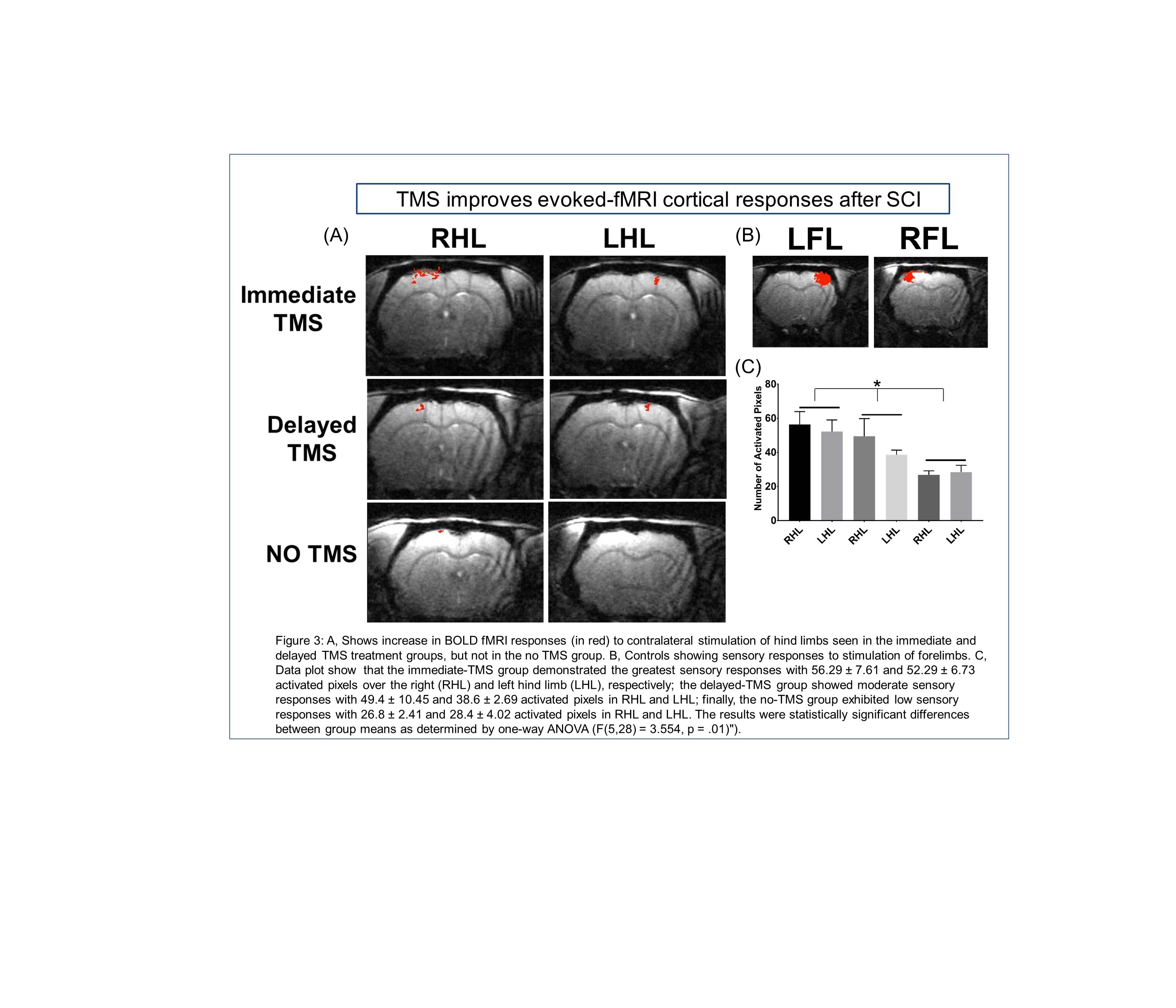
Results show that the immediate-TMS group demonstrated the greatest sensory responses with 56.29 ± 7.61 and 52.29 ± 6.73 activated pixels over the right (RHL) and left hind limb (LHL), respectively; the delayed-TMS group showed moderate sensory responses with 49.4 ± 10.45 and 38.6 ± 2.69 activated pixels in RHL and LHL; finally, the no-TMS group exhibited low sensory responses with 26.8 ± 2.41 and 28.4 ± 4.02 activated pixels in RHL and LHL, respectively. Motor behavior was assessed by weekly grid walk test, which indicated that the immediate-TMS group had fewer number of footfall errors indicating fastest recovery compared to the delayed-TMS and no-TMS groups but they all reached the same level at the end of the treatment.Discussion
Based on previously published evidence from our lab that verified the efficacy of TMS in improving outcome in traumatic brain injury, we sought to extend this successful outcome in the treatment of SCI. Our findings in this study indicate that TMS treatment improved cortical responses during fMRI. This indicates the improvement in the integrity of the corticospinal tract information. The results from the gridwalk indicated improvement in the test initially amongst the immediate TMS group, but then plateau together. Though the gridwalk is the gold standard in testing function and recovery in chronic SCI, it is not a sensitive test. Therefore there is a need to augment the data from the fMRI with more sensitive tests such as the motor evoked potential test. This will provide a cleaner perspective of the correlation of TMS treatment with recovery in SCI. We anticipate that application of TMS as a therapeutic strategy could be readily translated into the clinical setting as an alternative or adjuvant to traditional rehabilitation strategies.Acknowledgements
No acknowledgement found.References
Lu, Hongyang, et al. "Transcranial magnetic stimulation facilitates neurorehabilitation after pediatric traumatic brain injury." Scientific reports 5 (2015).Figures
TMS
improves evoked-fMRI cortical responses after SCI