5362
Representation of Taxonomic and Thematic Knowledge of the Human Brain1National Key Laboratory of Cognitive Neuroscience and Learning & IDG/McGovern Institute for Brain Research, Beijing Normal University, Beijing, People's Republic of China, 2Center for MRI research, Peking University, Beijing, People's Republic of China, 3MR Collaboration NE Asia, Siemens Healthcare, Beijing, People's Republic of China, 4Siemens Healthcare, Erlangen, Germany
Synopsis
Decades of studies have identified a list of
brain areas specific to a certain taxonomic category. However, neural
representations incorporating both taxonomic and thematic knowledge are not
well understood. In this study, we applied representational similarity analyses
to investigate the underlying organizing principles of high-resolution neural
activation patterns induced by different categories and themes at different
cortical levels. In contrast to taxonomic representation, we did not find
specific neural substrates representing thematic knowledge. Instead, neural
activation patterns specific to thematic information emerged only when
taxonomic differences were controlled for. These results suggest that the brain
is dominated by taxonomic knowledge and then modified by thematic knowledge.
Introduction
For a given entity, we know the category to which it belongs (e.g., an animal, a tool, etc.) and its associated context or location (e.g., in a sport metting, in a medical situation, etc.). Theories on how the brain represents aspects of conceptual knowledge are currently controversial as different studies have attributed different representations of conceptual knowledge to different brain regions, i.e. there is not yet a consensus on which brain region is responsible for which conceptual representation [1]. Such controversies are difficult to resolve using traditional paradigms (e.g., semantic priming and word or picture associations) because of potential confounding factors. In this study we employed a representation similarity analysis (RSA) approach using a set of stimuli whose taxonomic and thematic relationships were orthogonally manipulated to test the neural underpinnings of both the taxonomic and thematic aspects of different conceptual representations.Methods
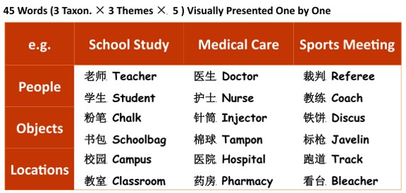
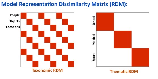
In this ER-designed fMRI study (N = 20), all participants (10 males; aged 18-27 years) were scanned on a MAGNETOM Prisma 3T MR scanner (Siemens, Erlangen, Germany) with a 20-channel head-neck coil. Forty-five printed words (Figure 1) belonging to 9 conditions [3 themes (school, healthcare, and sports) × 3 taxonomies (people, objects, and location)] were pseudo-randomly presented once in each run. Participants made taxonomic judgments in half of the 10 runs and thematic judgments in the other half so that they would have to explicitly access both the taxonomic and thematic aspects of each word. BOLD data were acquired using a prototype simultaneous multi-slice EPI sequence with the following parameters: number of slices=64, TR/TE=2000ms/30ms, FA=90º, slices acceleration factor =2, voxel size= 2×2×2mm3. The fMRI data were post-processed with SPM12 (the Wellcome Trust Center for Neuroimaging, http://www.fil.ion.ucl.ac.uk/spm/software/spm12/). We used a searchlight-based RSA [2] to evaluate voxel-wise activation patterns and detect the neural substrates generating these patterns. The activation pattern induced by each stimulus was correlated with model representational dissimilarity matrices grouped by taxonomic and thematic knowledge (Figure 2).Results
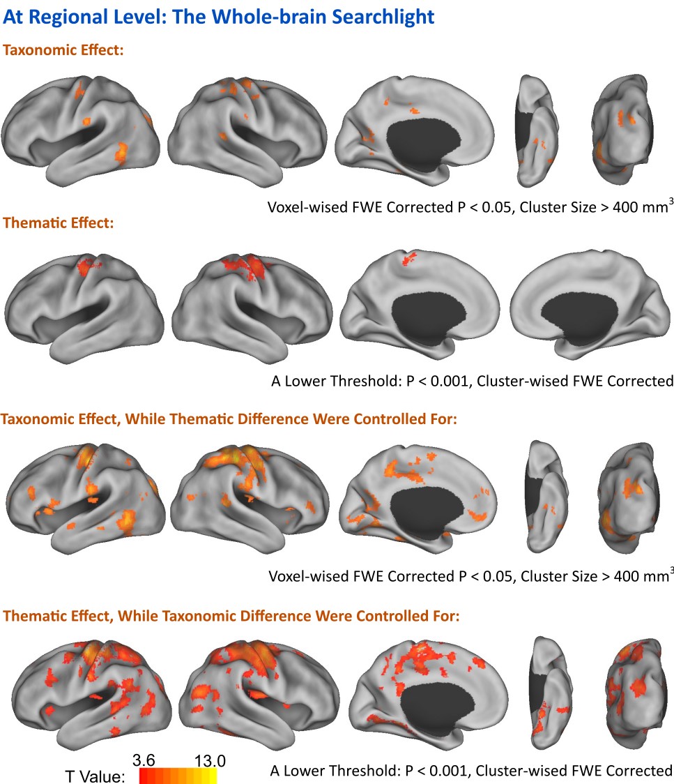
The results are presented in Figure 3. The following regions were found to represent taxonomic knowledge as they were significantly activated by particular taxonomic groupings of the stimuli: the left inferior frontal gyrus, the left transverse occipital sulcus, the left posterior temporal gyrus, the left parahippocampal gyrus, the bilateral retrosplenial cortices, and the left anterior superior temporal cortex. We did not find any brain region strictly representing thematic knowledge. However, when we did control for taxonomic differences, bilateral ventral temporal cortices, bilateral temporoparietal cortices, and the left inferior frontal gyrus were found to represent thematic knowledgeDiscussion and Conclusion.
Our results indicate that taxonomic and thematic knowledge share some of the same, but not all, neural substrates. While taxonomic knowledge is represented by category-specific regions, thematic information is represented in category-specific regions only when taxonomic differences are controlled for. These results suggest that taxonomic and thematic knowledge are not represented peer to peer, i.e., the brain is dominated by taxonomic knowledge and modified by thematic knowledge.Acknowledgements
Author contributions: YX and YB conceived and performed research, analyzed data and wrote the abstract; WM, TQ, TB and JG designed imaging sequence and helped collect imaging data.References
1 Jackson, R.L., et al. (2015) The nature and neural correlates of semantic association versus conceptual similarity. Cerebral Cortex 25, 4319-4333
2 Kriegeskorte, N., et al. (2008) Representational similarity analysis - connecting the branches of systems neuroscience. Front Syst Neurosci 2, 4
Figures