5361
Preliminary study of hypoxic exposure effect on pilots using the Resting-State Functional Magnetic Resonance Imaging1first affiliated hospital of zhengzhou university, zhengzhou, People's Republic of China
Synopsis
The study was about the Resting-State fMRI of pilots before and after hypoxic exposure. It proved that the feasibility of monitor the real-time cerebral functional activity change by using MRI. Otherwise, it confirmed that hypoxic exposure inhibited the cognitive functions of pilots to some extent.
Target audience
Radiologist and MR researchers, who interested in cerebral hemodynamic under conditions.
Purpose
To investigate how the cerebral function of resting-state changed in pilots with hypoxic exposure by using approaches of a fractional amplitude of low frequency fluctuation (fALFF) and regional homogeneity (ReHo).Introduction
Pilots often need to face and overcome all kinds of extreme situations during a flight, especially at upper atmosphere, where hypoxia is common due to low oxygen concentration and low pressure which cannot be fully avoided no matter how good of equipment sealing in modern aviation. As we all know, oxygen is indispensable for metabolism. In practice, pilots are often lack of oxygen unconsciously which may endanger the flight safety and cause the accident. In the past, we often used Ergonomic (Neuropsychological Scale), electroencephalogram and event related potential (ERP) to evaluate the cognitive function during acute hypoxia. In the current study, we used the resting-state blood oxygen level dependent functional magnetic resonance imaging (BOLD-fMRI) technology and two kinds of analysis methods (namely, fraction amplitude of low frequency (fALFF) and regional consistency (ReHo) ) to investigate the basic brain activity change after hypoxic exposure, and then fixed the corresponding brain areas.Methods
30 healthy male pilots (mean age 30 years, mean total flight time 1328 h) were included in this study. In order to investigate the change of the resting-state brain function in the condition of hypoxic exposure, the low oxygen mixed gas inhaled by participants through a breathing mask was approximate to the air composition at the altitude of 3000 m with the oxygen concentration of 14.5%. Pulse oximetry wasapplied to monitor in real-time the immediate pulse and oxygenation saturation ofeach subject pre- and post-hypoxic exposure. The subjects were instructed to keep as still as possible and not to think systematically. To reduce blurring and signal loss arising from field inhomogeneities, an automated high-order shimming method based on spiral acquisitions was used. Blood oxygen level dependent technique was appliedboth pre- (pre-OI) and post-low oxygen mixed gas inhalation (post-OI) using gradientEPI T2-weighted sequences, The EPI scan parameters were as follows: time of repetition(TR)=2,000ms; time of echo(TE)=35ms; flip angle=90°; field of vision=24cmx 24cm; matrix=64x64; slice thickness=4mm; scan time=410s. Amongthe tow times resting-state fMRI scanning using a 3D-FSPGR BRAVO sequence (GEMR750, WI, US) with the scanning parameters: TR/TE= 4632ms/10.5ms; FOV=24cm2; slice thickness = 4.0mm; bandwidth=62.5KHz; flip angle=111°.Thereafter, the fALFF and ReHo map data from two conditions were standardizedusing SPM8 software and paired t test was used for independent analysis. The threshold level was set at P<0.05 (FDR correction). Only the areas with more than 20 continuous voxels were sonsidered as effectively activated. We used XjView software to locate the anatomical locations. The results were shown by MRIcro softwear.Results
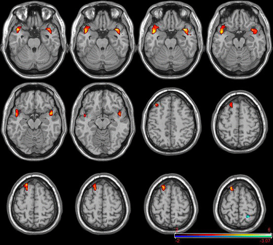
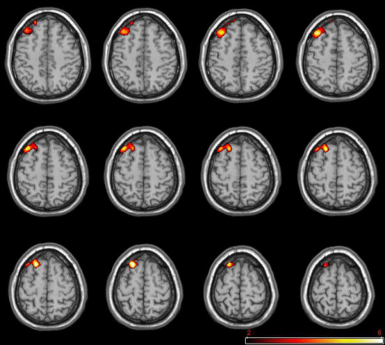
After hypoxic exposure, the pulse was (63.97±10.57) beats/min, the oxygen saturation was (92.37±3.85) %, it’s significant lower than initiate examination ((71.40±10.85) beats/ min, (96.27±1.28) %). Compared with pre-OI, significantly decreased fALFF post-OI was showed in various regions, including bilateral superior temporal gyrus(STG), the right superior frontal gyrus(SFG), while the left precuneus showed significantly increased fALFF. Meanwhile, ReHo values post-OI displayed the decreased regional statistics in the right superior frontal gyrus(SFG).Discussion
After hypoxic exposure, the oxygen saturation was kept around 92%, which was in the state of acute mild hypoxia1. Cahill et al.2-3 showed BOLD-fMRI signal of rats under hypoxic experiment was decreased comparing with the normal control. Both fALFF and ReHo values post-OI showed significantly decreased in SFG, while the left precuneus showed significantly increased fALFF. The two brain regions showed lower fALFF after hypoxic exposure demonstrated that hypoxia inhibits thecognitive functions of pilots to some extent. Some researchers4 used regional homogeneity (ReHo) approach to evaluate the resting-state brain function features of carbon monoxide poisoning patients. They found that the precunues had lower value of ReHo compared with control group, which was different with our research. We speculated that it may be relevant to the different composition of the gas, subjects and the analytical methods. The mechanism of the fALFF method needs to be further studied.Conclusion
The gradient EPI T2-weighted sequences scanning could monitor the resting-state cerebral activity changes in pilots with hypoxic exposure, and different approaches may demonstrate different results. Both the fALFF and ReHo values decreased significantly in SFG (P<0.05).Acknowledgements
First, I would like to thanks my parents for allowing me to realize my own potential. thanks for giving me all the support over the years. Next, I need to thank my supervisor and all the teachers encouraging me to pursue an advanced degree. I would also like to thank my friends who always took time to listen even when I was just complaining. Finally, I would like to thank my volunteers and all the people who help me overcome the difficulty and finish my research work.
References
1. Ernsting J. Aviat Space Environ Med, 1984, 55(3): 407-410;
2. Cahill LS, et al. Cereb Blood Flow Metab, 2014, 34(6): 1082-1088;
3. A Sørensen, et al. Ultrasound Obstet Gynecol, 2011, 38(6): 665-672;
4. Liu DH, et al. Cell Biochem Biophys, 2013; 67(3):1029-1032
Figures