5360
BOLD-fMRI evaluation of analgesic effects on allodynia-specific pain using fibromyalgia model rats1Department of Structural BioImaging, Faculty of Life Sciences, Kumamoto University, Kumamoto, Japan, 2Bruker BioSpin K.K., Yokohama, Japan
Synopsis
The aim of this study is to evaluate the effects of analgesic agents on the allodynia-specific response in an animal model of fibromyalgia. Before and after the treatment with analgesic agents, BOLD experiments using green laser stimulation were performed. Before the treatments with analgesic agents, S1, IC, and TH exhibited BOLD responses (S1: 1.1%, IC: 0.8%, TH: 0.7%). These responses were inhibited by pregabalin treatment and to a lesser extent by duloxetine treatment (S1: 0.4%, IC: not detected, TH: 0.4%). Our experimental system provides a robust preclinical and clinical evaluation system for new analgesic agents.
Introduction
Fibromyalgia and neuropathic disorders characterized by chronic pain induce the pathological condition “allodynia”, in which a stimulus that is normally not painful causes pain sensations. Allodynia is thought to develop by the potentiation of pain signals at the Aδ- and C-fibers [1]. Until now, the pain was evaluated by a patient-subjective assessment, using a visual analog score in humans, and by behavior tests in animals. The differences in the pain assessment methods make it difficult to smoothly transfer a preclinical study to a clinical study. Pain-mediated brain activation occurs in common in humans and animals, and thus it likely to be useful as an index of pain evaluation. The establishment of a pain evaluation system using BOLD-fMRI will contribute to the evaluation and development of new analgesic agents.
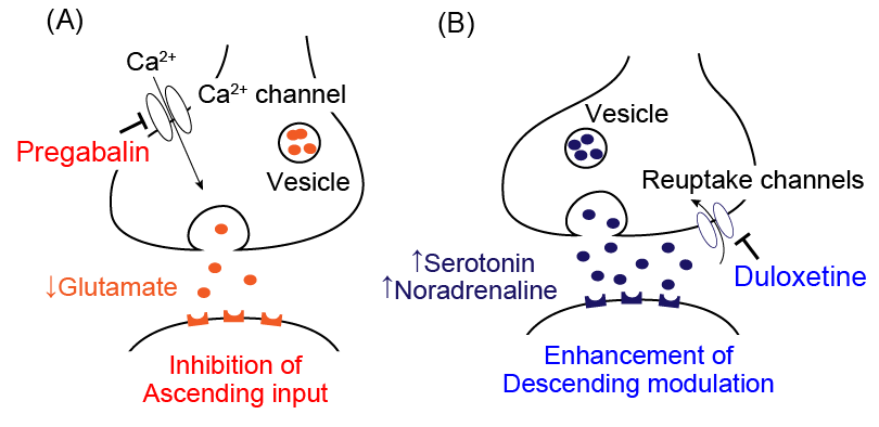
The aim of this study is to evaluate the effects of analgesic agents on the allodynia-specific response in an animal model of fibromyalgia, using BOLD-fMRI. We previously reported the suppression of allodynia-specific pain responses by pregabalin, using the 532 nm green laser [3]. In this study, we attempted to evaluate the analgesic effect of duloxetine, a treatment for fibromyalgia with different mechanisms from those of pregabalin (Fig. 1).
Methods
The reserpine-induced myalgia (RIM) rats, an animal model of fibromyalgia with the symptoms of allodynia, were converted according to the reported method [2]. Five RIM rats were used for the pregabalin experiments and three RIM rats were used for the duloxetine experiments. The rats were ventilated in a 70/30 mixture of N2/O2, and treated with gallamine and urethane (1.25 g/kg i.p.). Saline was administered to the rats (10 mL/kg i.v.), and 30 minutes later the BOLD experiments were performed. MRI experiments were performed with a 7.0 Tesla BioSpec 70/20 scanner and a rat brain 4-channel phased array surface coil (Bruker BioSpin). Functional data were acquired with a 4-shot GRE-EPI sequence (TR: 500 ms, TE: 15 ms, FA: 45°, matrix: 64 × 64, FOV: 2.56 × 2.56 cm2, 13 slices, slice thickness: 0.6 mm). The green laser with 350 mW output power and 2 s irradiation time was used to irradiate the left hind paws 5 times every 2 minutes, during the scans. After the first BOLD experiment, the rats were treated with pregabalin (10 mg/kg i.v.) or duloxetine (3 mg/kg i.v.), and 30 minutes later the same BOLD experiments were performed. The Independent Component Analysis (ICA) was performed with the FSL software. The brain regions that showed periodic BOLD responses with the frequency of the laser stimulation (8.3 mHz) were searched. The BOLD signal intensity was analyzed with the SPM8 software.Results
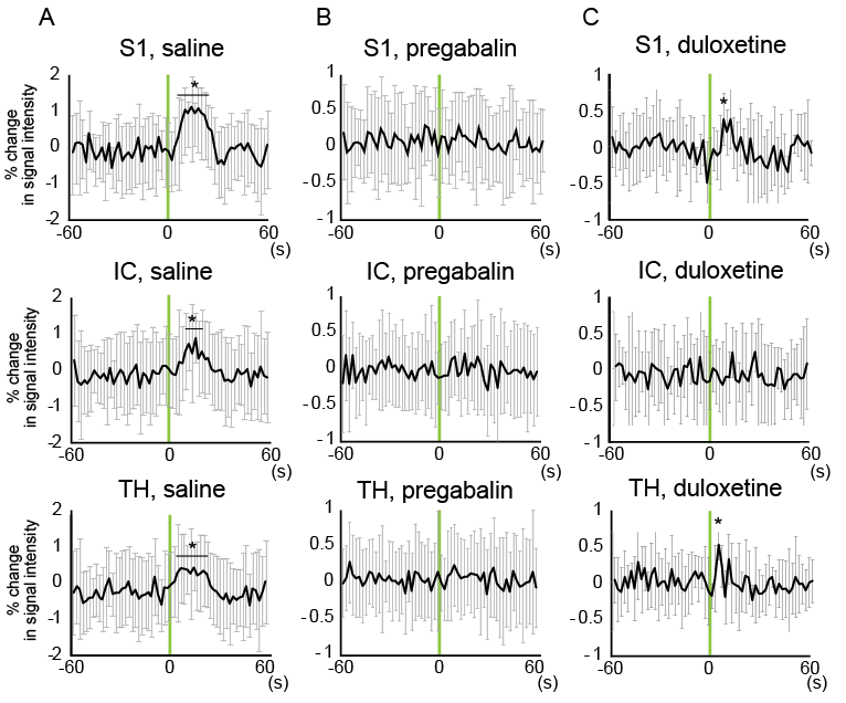
Before the administration of analgesic agents, through the ICA, the primary somatosensory cortex (S1), insular cortex (IC), and thalamus (TH) were found to exhibit periodic activation with the frequency of the laser stimulation (8.3 mHz). Upon the laser stimulation, the BOLD signal intensities in the S1, IC, and TH were increased by up to 1.1%, 0.8%, and 0.7%, respectively (Fig. 2A). When the rats were treated with pregabalin, no periodic activation with 8.3 mHz was detected by the ICA, and no significant BOLD signals were detected in the S1, IC, and TH, which had exhibited BOLD responses before the pregabalin treatment (Fig. 2B). When the rats were treated with duloxetine, the S1, IC, and TH signals were found to exhibit periodic activation with 8.3 mHz in 3 rats, 1 rat, and 2 rats, respectively. The BOLD signal intensities in the S1 and TH were increased by up to 0.4%. However, no significant BOLD signal changes were observed in the IC (Fig. 2C).Discussion
Before the treatments of analgesic agents, the S1, IC, and TH regions were activated by the green laser stimulation. S1 is the brain region related to pain sensation, and IC and TH are brain regions related to emotion caused by pain. Pregabalin reportedly binds to the voltage-dependent calcium channel, and suppresses the ascension of pain input. Thus, the BOLD responses in S1, IC, and TH were inhibited. It is conceivable that duloxetine mainly suppresses the BOLD response of IC by enhancing the descending pain modulation system and/or exerting an anti-depressive effect on IC. The discrimination of the brain responses between the pregabalin and duloxetine administrations indicates that our experimental system is applicable for the evaluation of the effects caused by different analgesic agents.Conclusions
We successfully observed the effects of fixed doses of pregabalin and duloxetine on the allodynia-specific responses. Our experimental system provides a robust preclinical and clinical evaluation system for new analgesic agents.Acknowledgements
We gratefully acknowledge Drs. Sokichi Honda, Keisuke Tamaki, Toshihiro Sekizawa, Masayasu Takahashi, Akihiko Fujikawa, and Shuichiro Kakimoto for fruitful discussions.References
[1] Ueda, H., Pharmacol. Ther. 109, 57–77 (2006), [2] Nagakura, Y. et al., PAIN 146, 26–33 (2009), [3] Yuzuriha, N. et al., Proc. Intl. Soc. Mag. Reson. Med. 24, 1722 (2016)
Figures