5359
Characteristics of brain spontaneous neuronal activity in chronic alcoholics using different resting-state fMRI algorithm1Radiology, Renmin Hospital of Wuhan University, Wuhan, People's Republic of China, 2GE Healthcare, GE MR Research China, People's Republic of China
Synopsis
Resting-state fMRI reflected spontaneous baseline neuronal activity.The fractional amplitude of low frequency fluctuation (fALFF) and regional homogeneity(ReHo) method had been developed to analyze the blood oxygenation level-dependent signal fluctuations in voxelwise analysis across the whole brain.In this study, we combined two resting-state fMRI algorithm to explore the features of brain spontaneous activities in chronic alcoholics.The results indicated the abnormality activities of some nodes in the default mode network and reward circuit. It is our hope that in future studies this technique may provide the opportunity to examine the integrity of networks involving the above loops in chronic alcoholics.
PURPOSE
Excessive alcohol abuse can cause structural and functional abnormalities of the brain1,2. Many methods have been proposed to assess alcohol-related brain damage with single algorithm.The present study used different resting-state fMRI algorithm(fALFF3 and Reho4) to detect the characteristics of spontaneous neuronal activity in chronic alcoholics,and to study the relationship between of resting state and the observed clinical symptom in chronic alcoholics.METHODS
24 alcohol dependent individuals and 22 healthy subjects were enrolled. They all underwent a resting state fMRI scanning on a GE 3.0T MR scanner. All resting state fMRI data were processed using DPARSF software based on MATLABE, SPM and REST operating environment to achieve the whole brain fractional amplitude of low frequency fluctuation (fALFF) data and the regional homogeneity (ReHo) data. One-sample t-tests were performed to analyze the difference of fALFF data and ReHo data within the alcohol dependent group and the healthy control group respectively. Two-sample t-tests were performed to analyze the difference between fALFF data and ReHo data of the two groups of the alcohol dependent group and the normal control group. The Montreal Neurological Institute (MNI) coordinates of the statistically significant region were recorded to achieve the specific anatomical location of these regions, with t recorded.RESULTS
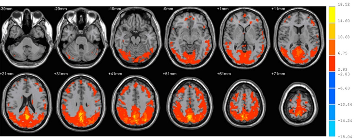
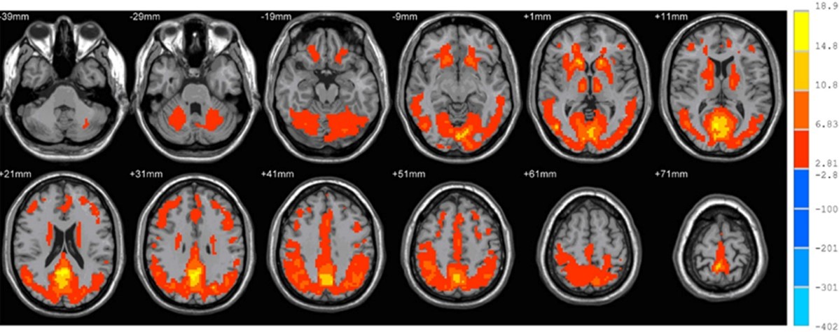
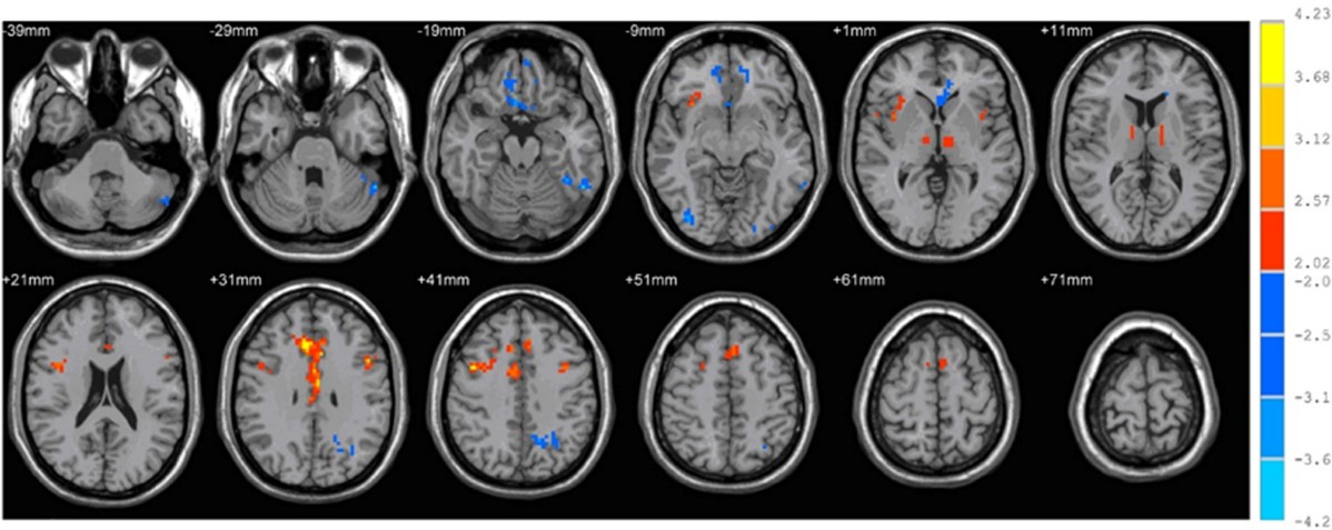
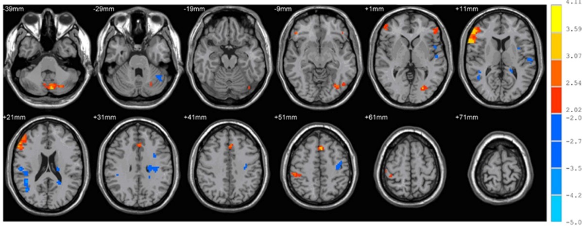
one-sample t-test results of fALFF within normal control group and alcohol dependent group indicated that fALFF values increased significantly in posterior cingulate cortex (PCC) and precuneus (PCu). Those areas were at the center, bilateral frontal, parietal, temporal lobe and anterior cingulate of the two groups (Fig.1);increased ReHo values were observed in the same regions where fALFF were increased and several additional areas included bilateral basal ganglia area and, cerebellum in both groups (Fig.2).Compared with the control group, the alcohol dependent group demonstrated that fALFF were reduced in bilateral medial prefrontal gyrus, left inferior temporal gyrus, left posterior lobe of cerebellum, right inferior occipital gyrus and left precuneus (P﹤0.05) (Fig.3), but increased in bilateral dorsal thalamus, anterior cingulate, bilateral inferior frontal gyrus, right middle frontal gyrus (P﹤0.05) (Fig.3).The alcohol dependent group demonstrated that ReHo were reduced in right superior temporal gyrus, bilateral postcentral gyrus, left posterior lobe of cerebellum, left insular lobe, left precentral gyrus, left caudate nucleus, the splenium of corpus callosum (P﹤0.05) (Fig.4), but increased in right postcentral gyrus, right middle and inferior frontal gyrus, left posterior lobe of cerebellum, left middle frontal gyrus, left inferior occipital gyrus and anterior cingulate (P <0.05) (Fig.5).CONCLUSIONS
With different resting-state fMRI algorithm,it indicated consistently that:1) Default mode network exists in alcohol dependent individuals, and the activity of the node of cerebellum and, precuneus are abnormal in alcohol dependent group. 2) Alcohol dependent individuals has abnormal activity in multiple brain areas during resting state, and these areas might be related with clinical manifestation and pathophysiology. 3) Similar to individuals with other substance addiction, reward circuit exist and the connection enhanced in alcohol dependent individuals.Acknowledgements
The authors would like to thank the anonymous reviewers for their significant and constructive comments and suggestions which greatly improved the paper. We are also gratefully thankful for the support and assistance from Hui Lin of the Advanced Application Team of GE Healthcare China.References
1.Harper, C., The neuropathology of alcohol-related brain damage. Alcohol Alcohol.2009;44(2):136-140.
2.Chanraud S,Pitel AL,Rohlfing T., et al., Dual tasking and working memory in alcoholism: relation to frontocerebellar circuitry. Neuropsychopharmacology.2010;35(9):1868-1878.
3.Zou QH,Zhu CZ.Yang Y., et al., An improved approach to detection of amplitude of low-frequency fluctuation (ALFF) for resting-state fMRI: fractional ALFF. J Neurosci Methods.2008;172(1): 137-141.
4.Zang Y,Jiang T,Lu Y., et al.,Regional homogeneity approach to fMRI data analysis. Neuroimage.2004;22(1):394-400.
Figures