5357
Working memory improved by abacus training in Chinese children: an fMRI study utilizing a spatial n-back task1Department of Physics, Zhejiang University, Hangzhou, People's Republic of China, 2Department of Psychology, Zhejiang University, Hangzhou, People's Republic of China
Synopsis
To examine whether abacus training improves working memory (WM), sixty-four children were randomly assigned into two groups, matched for intelligence. One group received abacus training for five years while the other group had no any abacus experience. WM was measured by a n-back task. The results showed that children with training were more accurate and faster than their peers. They also had greater activation and functional connectivity in the frontoparietal regions. The findings suggest that AMC training may be an effective method to improve WM in school children, which may have implications to help individuals with cognitive deficits.
Introduction
Working
memory (WM), the ability to temporarily maintain and manipulate goal-relevant
information, is critical for a wide range of cognitive activities, such as
reasoning and academic achievements[1]. It can be improved by training,
but the tasks used for training are often traditional WM paradigms that are far
from our daily life[2,3]. Such training may have difficulties in
generalizing to practical use. Recently, abacus-based mental calculation (AMC),
a method to improve arithmetical skills in Asian schools, has received much
attention. Given that the frontoparietal regions are critical for both AMC[4,5] and WM[6], this study investigated whether AMC training could improve
WM and alter underlying neural correlates in Chinese school children.
Method
Sixty-four children were recruited and then randomly assigned into AMC and control groups. The AMC group received two hours per week of AMC training from the first to the sixth grade while the control group was provided with a similar amount of extra learning using supplemental materials. In the sixth grade, their intelligence was examined by a raven test, and WM was assessed by a spatial n-back task (0-back, 2-back and 3-back). Neural activity during the n-back task was examined using fMRI. Participants with prolonged absence from the training or excessive head motion during fMRI scanning were excluded, resulting in 26 children (12 boys, mean age = 11.93 ± 0.54) in the AMC group and 22 children (10 boys, mean age = 11.92 ± 0.52) in the control group.Result
1. There was no group difference on intelligence (t(46)=0.88, p = 0.38).
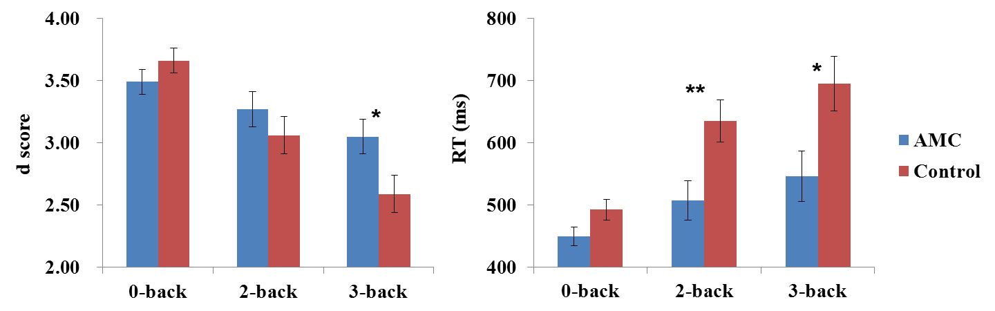
2. For d-prime score of the n-back task, a repeated measure ANOVA with Load (0-back, 2-back or 3-back) and Group (AMC or control) as factors indicated that there was an interaction between Load and Group (F (2, 92) = 4.81, p = 0.01). Post-hoc tests showed that children with AMC training had higher d scores than their peers in the 3-back condition (p < 0.03), but not in other conditions (Figure 1). A parallel repeated measure ANOVA on reaction time (RT) also revealed an interaction between Load and Group (F (2, 92) = 3.23, p < 0.05). Post-hoc tests showed that children with AMC training had shorter RTs than their peers in the 2-back (p < 0.01) and 3-back conditions (p = 0.02).
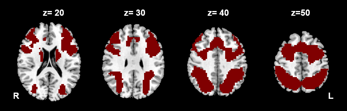
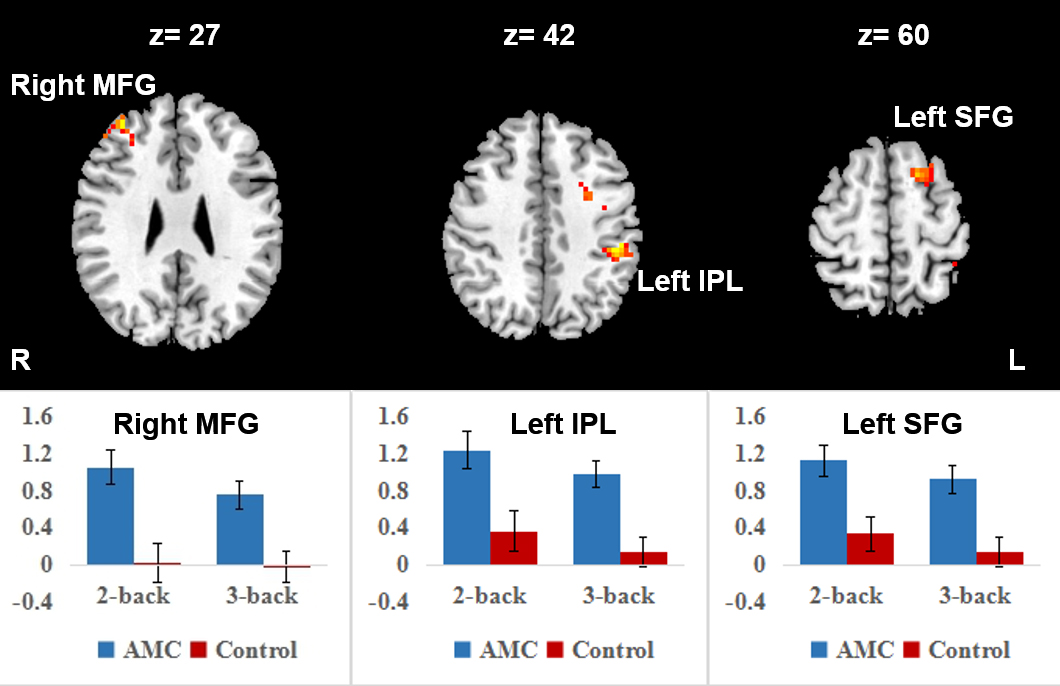
3. For the imaging data, two contrasts of interest were obtained for each individual: (1) 2-back vs. 0-back (2) 3-back vs. 0-back. To restrict the search space, the activation map for the 2-back and 3-back compared with the 0-back across all subjects was defined as a task mask (Figure 2). A factorial ANOVA with Load (2-back, 3-back) and Group (AMC or control) as factors was conducted. AlphaSim corrected significance level of p < 0.05 was obtained by a voxel-wise threshold of p < 0.01 and a minimal cluster size of 26 voxels. There was a main effect of Group in the right middle frontal gyrus (MFG), left inferior parietal lobule (IPL), and left superior frontal gyrus (SFG) (Figure 3). Children with AMC training had greater activation in these clusters than their peers. No clusters survived in the interaction between Load and Group.
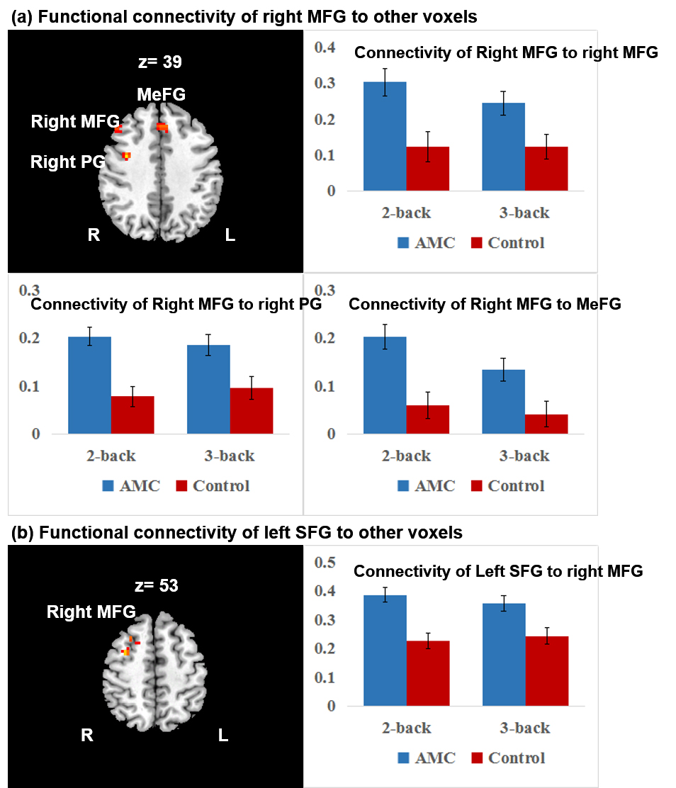
4. These clusters were used as seed regions to conduct seed-to-voxel functional connectivity. A parallel 2 (Load) × 2 (Group) factorial ANOVA showed a main effect of Group. Children with AMC training had greater functional connectivity between the right MFG and regions including the right precentral gyrus (PG), right MFG and medial frontal gyrus (MeFG), and between the left SFG and the right MFG (Figure 4). No clusters survived in the interaction between Load and Group.
Discussion
These findings suggest that long-term AMC training may improve WM and induce functional plasticity in the frontoparietal regions. It has been suggested that AMC relies on a visuospatial strategy4,5. Since the frontal-parietal network is associated with visual-spatial processing, it is possible that children with AMC training utilize more neural resources in frontoparietal regions and make these regions work more cooperatively. These findings may enhance our ability to treat WM deficits in the future.Acknowledgements
This study was supported by grants from the NSFC (No. 030900389 and No. 31270026).References
[1] Bull R, Espy KA, Wiebe SA (2008) Short-Term Memory, Working Memory, and Executive Functioning in Preschoolers: Longitudinal Predictors of Mathematical Achievement at Age 7 Years. Developmental neuropsychology 33: 205-228.
[2] Klingberg T, Fernell E, Olesen PJ, Johnson M, Gustafsson P, et al. (2005) Computerized Training of Working Memory in Children With ADHD-A Randomized, Controlled Trial. Journal of the American Academy of Child & Adolescent Psychiatry 44: 177-186.
[3] Diamond A, Lee K (2011) Interventions shown to aid executive function development in children 4 to 12 years old. Science 333: 959-964.
[4] Chen F, Hu Z, Zhao X, Wang R, Yang Z, et al. (2006) Neural correlates of serial abacus mental calculation in children: a functional MRI study. Neuroscience letters 403: 46-51.
[5] Hanakawa T, Honda M, Okada T, Fukuyama H, Shibasaki H (2003) Neural correlates underlying mental calculation in abacus experts: a functional magnetic resonance imaging study. NeuroImage 19: 296-307.
[6] Owen, A.M., McMillan, K.M., Laird, A.R., Bullmore, E. (2005) N-back working memory paradigm: A meta-analysis of normative functional neuroimaging studies. Human brain mapping, 25:46-59.
Figures