5354
Alteration of intrinsic brain activity in children with obstructive sleep apnea syndrome revealed by resting-state functional magnetic resonance imaging1Imaging Center, Beijing Children's Hospital, Capital Medical University, Beijing, People's Republic of China, 2The State Key Laboratory of Management and Control for Complex Systems, Institute of Automation, Chinese Academy of Sciences, Beijing, People's Republic of China, 3Research Center for Brain-inspired Intelligence, Institute of Automation, Chinese Academy of Sciences, Beijing, People's Republic of China, 4Center for Excellence in Brain Science and Intelligence Technology, Chinese Academy of Sciences, Beijing, People's Republic of China, 5Department of pattern recognition and intelligent system, University of Chinese Academy of Sciences, Beijing, People's Republic of China, 6Otolaryngology, Beijing Children's Hospital, Capital Medical University, Beijing, People's Republic of China, 7Neurology department, Beijing Children's Hospital, Capital Medical University, Beijing, People's Republic of China, 8MR Research China, GE Healthcare, Beijing, People's Republic of China
Synopsis
Obstructive sleep apnea syndrome (OSAS) in adults has been demonstrated tobe associated with brain functional and structural changes. However, little is known about the changes in regional synchronization of spontaneous brain activity and spontaneous fluctuations in children with OSAS. In the present study, regional homogeneity (ReHo) analysis and amplitude of low-frequency fluctuation (ALFF) based on resting-state functional magnetic resonance imaging (MRI) were used to investigate spontaneous brain activity in children with OSAS compared with controls (CN). As a result, children with OSAS showed significant functional alterations of the cerebellum and temporal gyrus in children with OSAS.
Purpose
Obstructive sleep apnea syndrome (OSAS) in adults has been demonstrated to be associated with brain functional and structural changes. However, little is known about the changes in regional synchronization of spontaneous brain activity and spontaneous fluctuations in children with OSAS. The purpose of the present study was to investigate spontaneous brain activity in children with OSAS compared with normal controls (CN) using regional homogeneity (ReHo) analysis and amplitude of low-frequency fluctuation (ALFF) based on resting-state functional MRI (R-fMRI).Materials and Methods
R-fMRI protocol
Eleven children with OSAS (1 females, mean age = 5.18, SD = 2.23,) and ten healthy controls (5 females, mean age = 7.30, SD = 2.21) were included in this study. Participants were scanned in a 3.0T GE Discovery MR750 scanner at Beijing Children’s Hospital. For each subject, R-fMRI and structural T1 images were acquired. In addition, clinical parameters including information of disease, whether taking medicine, intelligence and the information of sleep were recorded.
fMRI preprocessing
The preprocessing procedure using DPARSF and SPM8 toolboxes included removing the first 10 volumes, slice-timing correction, reoriented realigning and normalizing to the MNI space. Then smoothing (Gaussian kernel, 6 mmFWHM) and band-pass filtering (0.01-0.08Hz) were optional according to the following processing. In addition, the linear trend and 24 movement parameters resulting from rigid body correction for head motion were removed.
fMRI processing
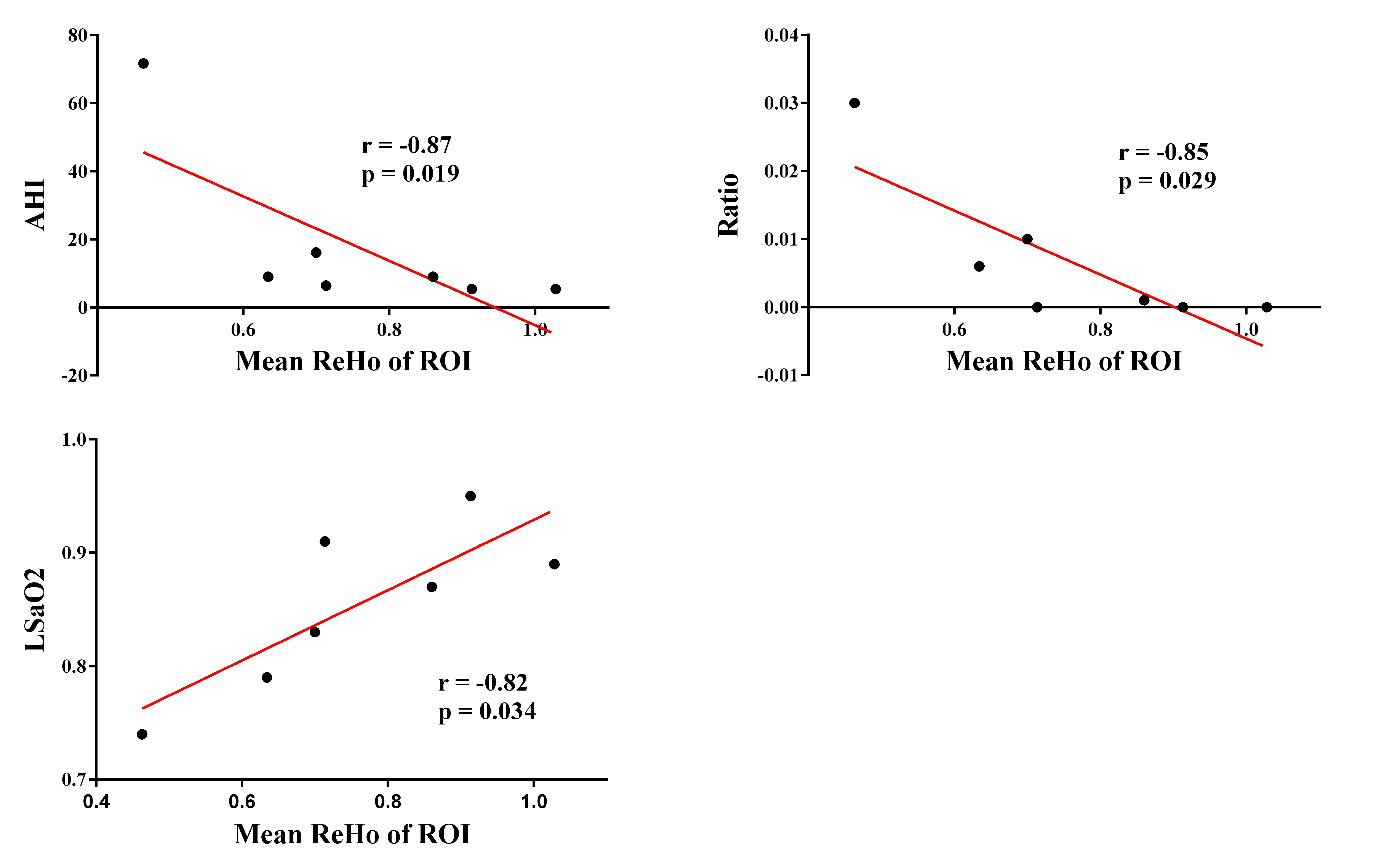
Two measures which are related to characteristic information of R-fMRI data were obtained including ReHo and ALFF.1,2 Two sample t-test with one tail was conducted to explore group-level difference. In addition, age, sex, disease course and whether or not taking medicine related to asleep were regressed out to reduce their effects. The significance threshold was p<0.001, topological FDR corrected. To explore the relationship between clinical parameters and ReHo or ALFF values of brain regions which have significant between group difference, the mean ReHo and ALFF values were extracted and correlated with those clinical parameters.
Result
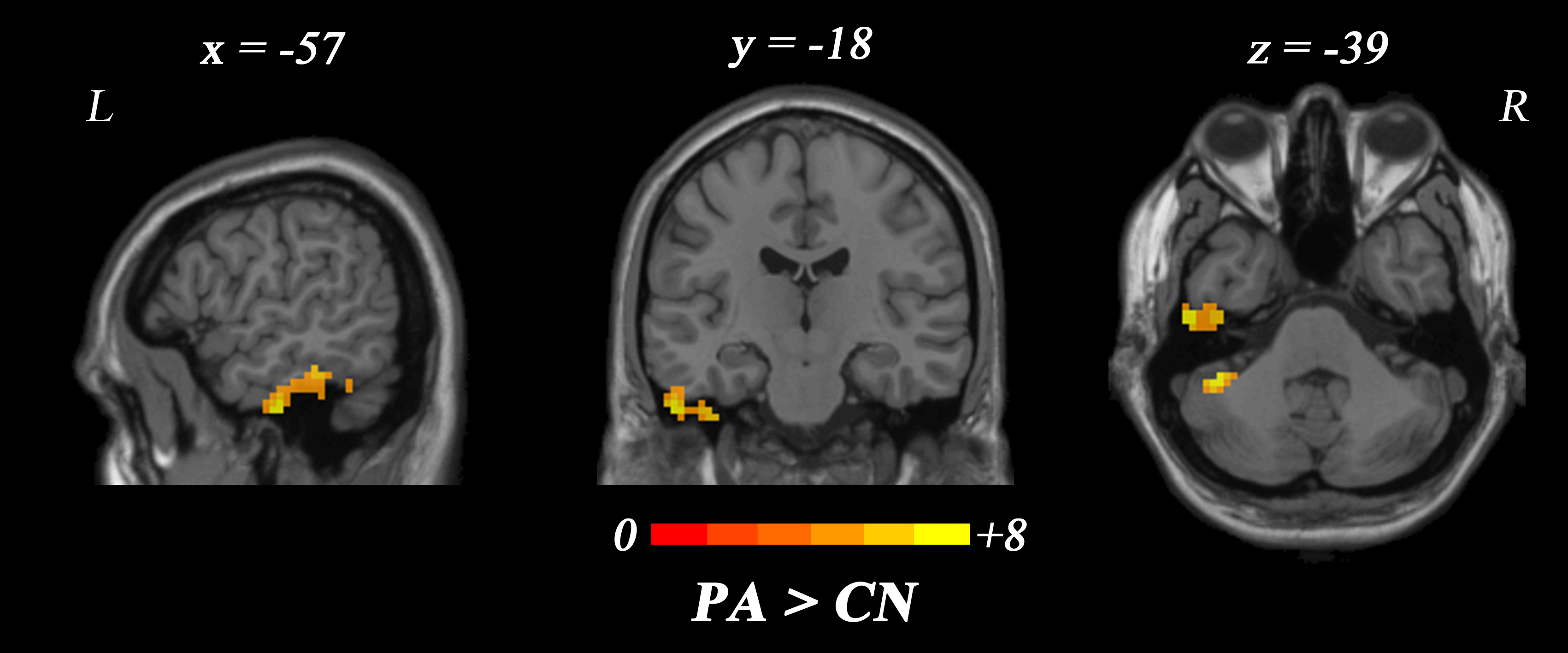
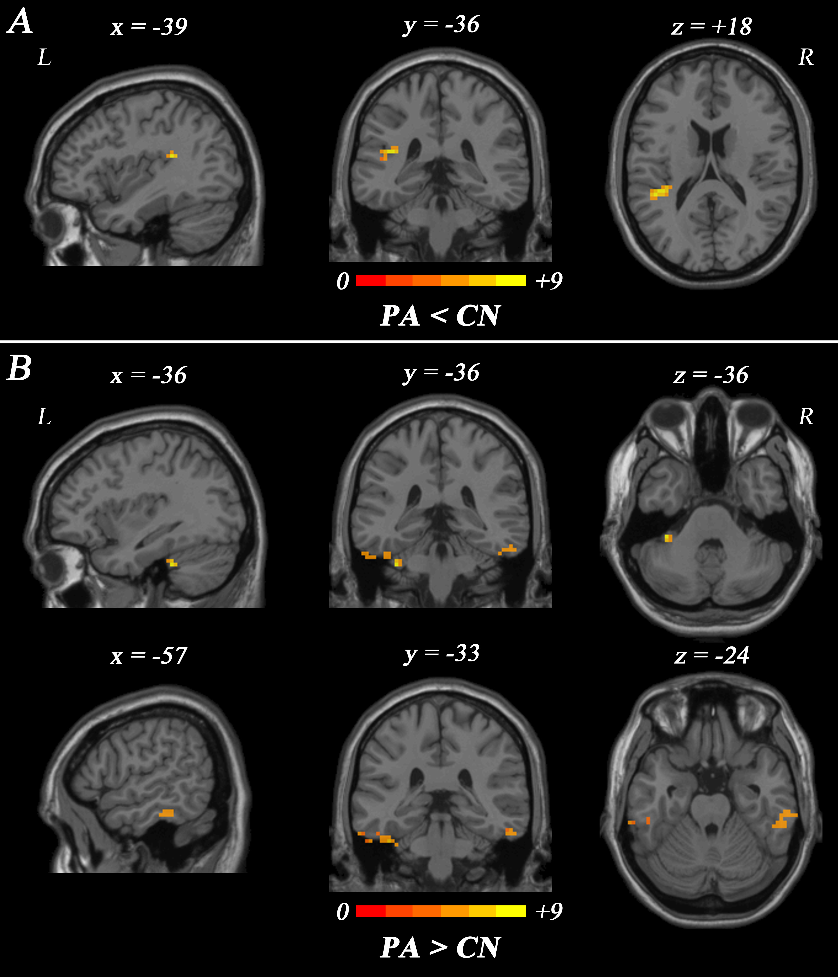
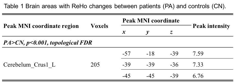
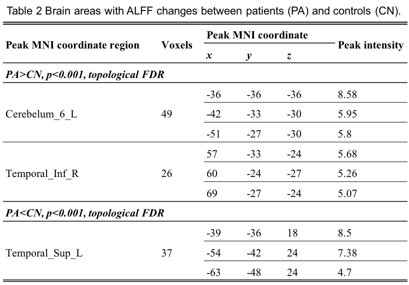
Compared to healthy controls, the value of ReHo on left cerebellum was significantly increased (Fig.1 and Table 1). In addition, the values of ALFF on left superior temporal gyrus were significantly increased and the values on left cerebellum and right inferior temporal gyrus were significantly decreased compared to normal controls (Fig.2 and Table 2). Besides, significant correlation between mean ReHo of ROI and AHI, MOS and Ratio were found in patient group (Fig.3). And correlation between mean ReHo and OAI, HI, sleep efficiency, or arousal index was not significant. At last, the correlation between mean ALFF values and those clinical parameters was also not significant.Discussion
ReHo analysis showed higher ReHo in the left cerebelum. ALFF analysis showed higher ALFF in the left cerebelum and the right in ferior temporal gyrus, while lower ALFF in the left superior temporal gyrus in pediatric OSAS patients. A recent study found that adult patients with OSAS had significant different ReHo values in distributed cortical and sub-cortical regions compared to CN.3 Those regions were different from our study. The reason for this phenomenon could be that functional alterations of children OSAS patients were different from those of adult patients, and also demonstrate that OSAS is not only an upper airway disease but may also be associated with brain functional alterations.Conclusion
Children with OSAS showed significant functional alterations in the cerebellum and temporal gyrus. The ReHo and ALFF methods are useful noninvasive tools for detection of early changes of brain function in children with OSAS.Acknowledgements
No acknowledgement found.References
1 Zang Y, Jiang T, Lu Y, et al. Regional homogeneity approach to fMRI data analysis. Neuroimage. 2004;22(1):394-400.
2 Yang H, Long XY, Yang Y, et al. Amplitude of low frequency fluctuation within visual areas revealed by resting-state functional MRI. Neuroimage. 2007;36(1):144-152.
3 Peng DC, Dai XJ, Gong HH, et al. Altered intrinsic regional brain activity in male patients with severe obstructive sleep apnea: a resting-state functional magnetic resonance imaging study. Neuropsychiatr Dis Treat. 2014;19(10):1819-1826.
Figures