5352
Acute effects of alcohol on brain perfusion monitored with 3D pseudo continuous arterial spin labeling(3D PCASL)liang zhang1 and jun chen2
1radiology, Renmin Hospital of Wuhan University, Wu Han, People's Republic of China, 2Radiology, Renmin Hospital of Wuhan University, Wu Han, People's Republic of China
Synopsis
Despite the fact that alcohol have impact on human brain function, the mechanism is not yet well understood. In order to detect the changes of blood perfusion information based on PCASL, and to discuss the possible mechanism, we analyze the differences of CBF in 29 healthy volunteers before and after alcohol consumption. The results show that CBF changed significantly in brainstem, bilateral hippocampus, left frontal lobe, bilateral thalamus, corpus callosum, anterior cingulate cortex, bilateral cerebellum, bilateral occipital lobe, cuneus and bilateral temporal gyrus. These abnormal activities may have important value in revealing the mechanism of alcohol in the brain.
Purpose
Alcohol may disrupt hemodynamic response and local nerve activity which impact brain function, but to date the mechanism is not fully clear. Utilizing whole brain 3D pseudo continuous arterial spin labeling (PCASL), we aimed to preliminary explore the hemodynamic impact of taking in alcohol by evaluating cerebral blood flow (CBF) change of healthy volunteers. This will helps us on a better understanding on the activities of brain function after drinking and mechanisms of the influences from alcohol on the corresponding brain areas.Method
This study was approved by the institutional review board, and informed consents were obtained from all subjects. In our study, 29 healthy volunteers (23 male,6 female, mean age of 25.03±2.67 years) consumed moderate doses of alcohol (male 0.6 g/kg, female 0.55 g/kg). MRI examinations were performed before and 30min after alcohol consumption on a GE Discovery MR750 Plus 3.0T superconducting magnetic resonance(GE Healthcare, Milwaukee, Wisconsin, USA) which was equipped with a 8-channel phased-array head coil. Conventional MRI sequences included General Axial T2WI and 3D PCASL using 3D spiral fast spin echo pulse sequence were acquired during scan. PCASL detailed parameters are: TR=4632ms, TE=10.5ms, NEX=3, 4mm thickness, 0mm interlayer spacing, 8 arms with 512 sampling points, FOV =24cm×24cm, 36 axial slices, Post Labeling Delay 1525ms. CBF maps are then obtained using functool post processing software on a GE AW workstation. SPM8 was applied in data processing for comparison of cerebral functioning before and after alcohol consumption. Pairing T-test was adopted to examine the differences of CBF.Results
Compared data of 30min after drinking with before-drinking: the CBF In brainstem, bilateral hippocampus, left frontal lobe, bilateral thalamus, corpus callosum, and anterior cingulate cortex were significantly increased in table 1, and bilateral cerebellum, bilateral occipital lobe, cuneus and bilateral temporal gyrus were significantly decreased in table 2. Figure 1 demonstrates the differences of CBF in the brain before after drinking(P<0.001, Alphasim corrected).Conclusion and discussion
The source of the ASL signal is related to neurophysiology[1], which can provide information related to brain activity, and cerebral blood flow (CBF) is a marker of neurological activity[2]. The difference of blood perfusion information was found in the brain,which indicated that there were potential local brain area abnormal functional activity exists after drinking. These changes may be associated with metabolic, hormonal dynamics and hemodynamic autoregulation, which may have interaction effects or influenced by other neural mechanisms .Acknowledgements
Grateful acknowledgement is made to Prof. Jun Chen, my tutor,who gave me considerable help by means of suggestion, comments and criticism. Without his pushing me ahead, the completion of this thesis would be impossible. In addition, I deeply appreciate the contribution to this thesis made in various ways by my colleagues Shihao Wu,Qizhong Xu,Peifu Li and Yawen Ao, who have generously offered their consistent support and encouragement.References
[1] Detre J A, Wang J. Technical aspects and utility of fMRI using BOLD and ASL[J]. Clin Neurophysiol,2002,113(5):621-634.[2] Rao H, Wang J, Tang K, et al. Imaging brain activity during natural vision using CASL perfusion fMRI[J]. Hum Brain Mapp,2007,28(7):593-601.Figures
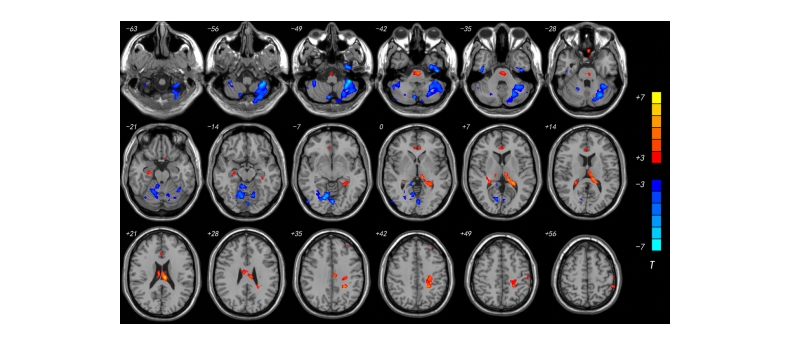
Fig.1 Differences in cerebral blood flow before and after drinking(P<0.001,Alphasim corrected).
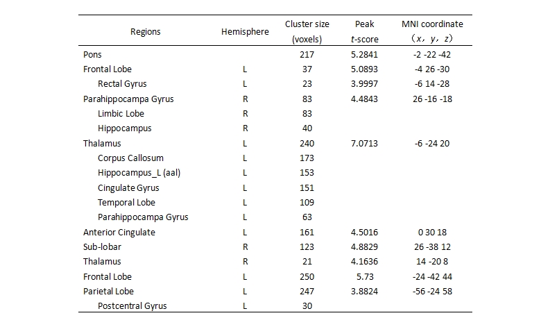
Tab.1 Peak MNI coordinates of the 13 clusters of the paired t-test result of before and after drinking-brain functional areas’ increased(P<0.001,Alphasim corrected)
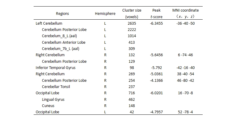
Tab.2 Peak MNI coordinates of the 13 clusters of the paired t-test result of before and after drinking-brain functional areas’ decreased(P<0.001,Alphasim corrected)