5346
Voxel-level comparison of regional neural activity in patients with type 2 diabetes mellitusDong Zhang1, Changzheng Shi1, Rong Ma1, Zhongping Zhang2, and Liangping Luo1
1Medical Imaging Center, The First Affiliated Hospital of Jinan University, Guangzhou, People's Republic of China, 2MR Research China, GE Healthcare, Beijing
Synopsis
People with high risk of diabetes exhibited decreased ReHo in right temporal lobes, left pallidum/ lenticular nucleus/insula/Heschl gyrus and left cingulate cortex and declined fALFF in left Heschl gyrus, left supramarginal gyrus and left cingulate cortex, which is consistent with type 2 diabetes mellitus (T2DM) patients. These abnormal regions may be regarded as the endophenotype of T2DM in resting-state BOLD-fMRI.
Introduction
We aimed to find the endophenotype of type 2 diabetes mellitus (T2DM) by investigating the regional neural activity patterns of T2DM population and their discordant siblings of first degree relatives without diabetes (high risk, HR group). Conventional resting-state blood oxygenation level dependent effect functional MRI (BOLD-fMRI) was used in this study.Methods
Twenty six pairs of T2DM patients and their discordant siblings of first degree relatives without diabetes were included in this study. In addition, twenty six age- and gender-matched healthy volunteers were also recruited as control group (HC group). All participants underwent anatomical MRI and resting-state BOLD-fMRI scans on a 3.0T whole-body MRI scanner (Discovery 750, GE Medical System, Milwaukee, WI). All BOLD-fMRI images were post-processed using SPM8 package in MATLAB (version 7.6). Typical resting-state fMRI metrics including Regional homogeneity (ReHo) and fractional amplitude of low frequency fluctuation (fALFF) were calculated and compared with HC group.Results
As compared with HC group, T2DM group showed decreased ReHo in right temporal lobes, left pallidum / lenticular nucleus / Heschl gyrus / insula and left cingulate cortex, while declined fALFF was observed mainly in bilateral lingual gyrus / precuneus, left cingulate cortex, left Heschl gyrus and left supramarginal gyrus. The HR group exhibited decreased ReHo in bilateral temporal lobes, left pallidum/ lenticular nucleus/insula/Heschl gyrus and left cingulate cortex. However, declined fALFF was observed mainly in left Heschl gyrus/insula, left supramarginal gyrus and left cingulate cortex.Discussion and Conclusion
The high risk population demonstrated decreased ReHo in right temporal lobes, left pallidum/ lenticular nucleus/insula/Heschl gyrus and left cingulate cortex in consistent with T2DM patients, and declined fALFF in left Heschl gyrus, left supramarginal gyrus and left cingulate cortex. These abnormal regions may be regarded as the endophenotype of T2DM in resting-state BOLD-fMRI.Acknowledgements
No acknowledgement found.References
No reference found.Figures
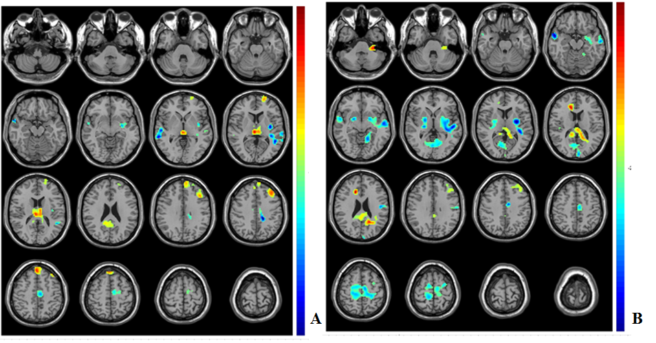
Altered
Reho
in T2DM patients (A) and the high-risk population (B) as compared with
healthy controls. Reho decreased in right temporal lobes, left pallidum/ lenticular
nucleus / insula / Heschl gyrus
and left cingulate cortex of both T2DM patients and high-risk
population.
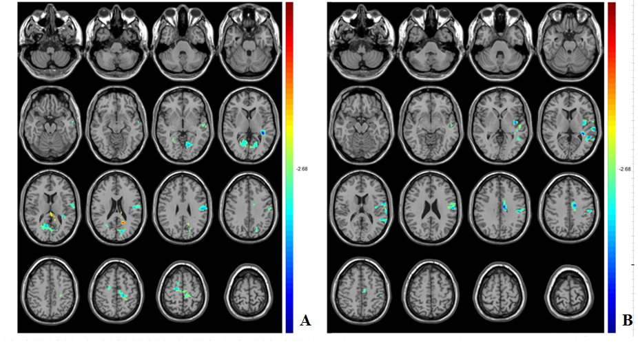
Altered
fALFF
in T2DM patients (A) and the high-risk population (B) as compared with
healthy controls. We observed the decline
of fALFF in left Heschl
gyrus, left supramarginal
gyrus and left cingulate cortex of both T2DM patients and high-risk
population.