5329
Investigating the feasibility of classifying independent components in resting state BOLD fMRI with sparse paradigm free mapping1Basque Center of Cognition, Brain and Language, Donostia - San Sebastian, Spain, 2Biodonostia Health Research Institute, Donostia-San Sebastian, Spain, 3Neurology Department, University Hospital Marqués de Valdecilla, Santander, Spain, 4Neuroscience Area, Biodonostia Health Research Institute, Donostia-San Sebastian, Spain, 5Centro de Investigacion Biomedicas en Red Enfermedades Neurodegenerativas (CIBERNED), Institute Carlos III, Spain, 6Ikerbasque. Basque Foundation for Science, Bilbao, Spain
Synopsis
This work proposes a novel method for the classification of ICs in resting-state fMRI data based on sparse paradigm free mapping (PFM), a deconvolution approach that enables detecting BOLD events without prior information of their timing. This approach uses a single temporal feature, the significance of the deconvolution model estimated with PFM. Our results demonstrate that despite its simplicity this approach achieves similar sensitivity in classifying the neuronal-related BOLD components to the more complex classification method of ICA-AROMA, but with less specificity in classifying noise components. In addition, it can improve the identification of physiological noise components.
Introduction
Several automated methods have been proposed for denoising of the BOLD fMRI signal based on the independent component analysis where the basic procedure is to classify the independent components (IC) as either neuronal-related BOLD or noise components and then remove the latter before reconstructing the dataset1-9. These methods attempt to overcome the limitations of a manual classification of the ICs10, which is a time consuming process, difficult to reproduce and requires expertise. Multiple temporal, spectral and spatial features can be computed from the IC time series and spatial maps (e.g. fraction of spectral power, correlation with realignment parameters, or extent of the IC map in ventricular CSF), where the number of features is highly variable across algorithms. In this work, we propose a novel and simple method based on sparse paradigm free mapping (PFM)11, a deconvolution approach that detects neuronal-related BOLD events without prior information of their timing, for the classification of ICs in resting-state fMRI data. This approach only uses a single feature, the statistical significance of the deconvolution estimated with PFM.Methods
Data acquisition and analysis: 79 subjects were scanned in a Siemens Trio 3T scanner with a 32 channel head coil during resting state while fixating eyes on a white cross (TR/TE: 2000/28 ms, FA: 78º, FOV: 192x192 mm, voxel size: 3x3x3 mm3, 33 slices). Datasets were corrected for head motion and spatially smoothed (5 mm FWHM Gaussian) with FSL (FMRIB, Oxford, UK). Subsequently, the preprocessed datasets were analyzed with probabilistic ICA (MELODIC)12 where the order of the decomposition was estimated based on the Laplace approximation (LAP) method.
ICA Classification: The basis of our classification approach is that if the IC is of neuronal origin, its time series must include neuronal-related BOLD events that can be detected by deconvolution of the IC time series with PFM11. In contrast, if the IC is related to noise or artefacts, the time series must not include any events, i.e. the deconvolved time series must be zero. Hence, for each subject, we computed the deconvolution of the IC time series with the PFM implementation in AFNI (see 3dPFM, https://afni.nimh.nih.gov/pub/dist/doc/program_help/3dPFM.html) using the SPM canonical HRF, the Dantzig Selector and the Bayesian Information Criterion. The normalized z-score of the F-statistic of a full model comprising the deconvolved BOLD events was also computed for each component. Our hypothesis is that a high z-scores (z > 0) indicates high probability of a neuronal-related BOLD IC, whereas a z-score equal to 0 is obtained when no events are detected. For evaluation, ICs were also classified manually10 and with the automated method ICA-AROMA3 with default parameters.
Results
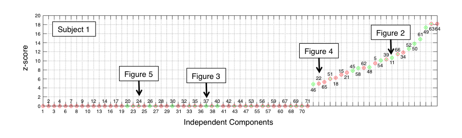
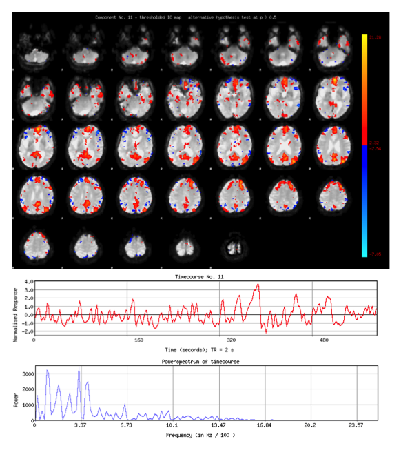
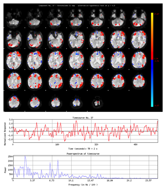
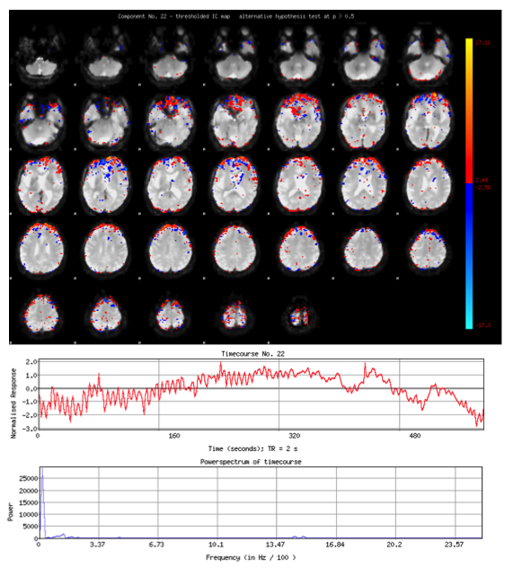
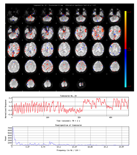
Figure 1 summarizes the results of the classifications, where the ICs are ordered with increasing z-scores, for an illustrative subject. Figure 2 shows the spatial maps, time courses and spectrum of IC#11, which was labeled as neural-related by the manual classification, the PFM-based approach and ICA-AROMA since it clearly represents the default mode network. Figure 3 shows IC#37, whose spatial map clearly indicates that the component is related to the dorsal attention network and it was correctly classified as neuronal-related in the manual and ICA-AROMA classification. However, this component was misclassified by the PFM-based approach (z = 0). Figure 4 shows IC#22, which was labeled as noise in the manual classification and by ICA-AROMA, whereas the PFM-based method classify it as neuronal-related (z > 0) despite it clearly represents a motion-related component with the largest amplitudes in the edges of the brain. Figure 5 shows IC#24, which was labeled as noise in the manual classification and the PFM-based approach, but as neuronal-related BOLD by ICA-AROMA despite its spatial map and spectrum clearly indicates it represents cardiac pulsations. Similar results were observed for the rest of subjects.Discussion and Conclusion
Our results indicate that the proposed simple classification rule based on the z-score of the PFM-deconvolved model achieves similar sensitivity to ICA-AROMA in classifying the neuronal-related BOLD components, but less specificity in distinguishing the noise components. They also suggest that using a temporal feature based on the deconvolution of the IC time course with PFM might help to improve the classification of certain components, mainly related to physiological fluctuations. This observation calls for the combination of the proposed method with additional temporal features, but mainly spatial features such as the smoothness or entropy of the IC map, or the extent of the IC map with the edges of the brain or ventricular CSF, in order to increase the specificity of correctly identifying motion-related components.Acknowledgements
This work was supported by the Spanish Ministry of Economy and Competitiveness [grant PSI 2013-42343 Neuroimagen Multimodal]; and the Severo Ochoa Programme for Centres/Units of Excellence in R&D [SEV-2015-490].References
[1] Bhaganagarapu, K., Jackson, G.D., Abbott, D.F., 2013. An automated method for identifying artifact in independent component analysis of resting-state fMRI. Front. Hum. Neurosci.. 7, 343.
[2] De Martino, F., Gentile, F., Esposito, F., Balsi, M., Di Salle, F., Goebel, R., Formisano, E., 2007. Classification of fMRI independent components using IC-fingerprints and support vector machine classifiers. Neuroimage 34(1), 177–194.
[3] Pruim, R.H., Mennes, M., van Rooij, D., Llera, A., Buitelaar, J.K., Beckmann, C.F., 2015a. ICA-AROMA: A robust ICA-based strategy for removing motion artifacts from fMRI data. Neuroimage 112, 267-277.
[4] Pruim, R.H., Mennes, M., Buitelaar, J.K., Beckmann, C.F., 2015b. Evaluation of ICA-AROMA and alternative strategies for motion artifact removal in resting state fMRI. Neuroimage 112, 278-287.
[5] Rummel, C., Verma, R.K., Schöpf, V., Abela, E., Hauf, M., Berruecos, J.F.Z., Wiest, R. 2013. Time course based artifact identification for independent components of resting-state fMRI. Front. Hum. Neurosci. 7, 214.
[6] Salimi-Khorshidi, G., Douaud, G., Beckmann, C.F., Glasser, M.F., Griffanti, L., Smith, S.M., 2014. Automatic denoising of functional MRI data: combining independent component analysis and hierarchical fusion of classifiers. Neuroimage 90, 449–468.
[7] Sochat, V., Supekar, K., Bustillo, J., Calhoun, V., Turner, J.A., Rubin, D.L., 2014. A robust classifier to distinguish noise from fMRI independent components. PLoS One 9, e95493.
[8] Tohka, J., Foerde, K., Aron, A.R., Tom, S.M., Toga, A.W., Poldrack. R.A., 2008. Automatic independent component labeling for artifact removal in fMRI. Neuroimage, 39, 1227–1245.
[9] Wang, Y., Li, T-Q., 2015. Dimensionality of ICA in resting-state fMRI investigated by feature optimized classification of independent components with SVM. Front. Hum. Neurosci. 9, 259.
[10] Kelly, R.E. Jr, Alexopoulos, G.S., Wang, Z., Gunning, F.M., Murphy, C.F., Morimoto, S.S., Kanellopoulos, D., Jia, Z., Lim, K.O., Hoptman, M.J., 2010. Visual inspection of independent components: defining a procedure for artifact removal from fMRI data. J. Neurosci. Methods 189, 233-45.
[11] Caballero-Gaudes, C., Petridou, N., Francis, S.T., Dryden, I.L., Gowland, P.A., 2013. Paradigm free mapping with sparse regression automatically detects single-trial functional magnetic resonance imaging blood oxygenation level dependent responses. Hum. Brain. Mapp. 34, 501-518.
[12] Beckmann, C.F., Smith, S.M., 2004. Probabilistic independent component analysis for functional magnetic resonance imaging. IEEE Trans Med Imaging. 23(2):137-52.
Figures