5317
Resting state connectivity analysis for normal and abnormal physiology conditions during fMRI data acquisition1Center for Neuroscience Imaging Research, Institute for Basic Science (IBS), Suwon, Korea, Republic of, 2Department of Health Sciences and Technology, SAIHST, Sungkyunkwan University, Seoul, Korea, Republic of, 3Center for Molecular and Cellular Imaging, Samsung Biomedical Research Institute, Seoul, Korea, Republic of, 4Department of Radiology, Samsung Medical Center, Sungkyunkwan University School of Medicine, Seoul, Korea, Republic of, 5Department of Electronic, Electrical and Computer Engineering, Sungkyunkwan University, Suwon, Korea, Republic of
Synopsis
Unlike blood oxygen level dependent (BOLD)-fMRI, accurate physiologic adjustment for subjects during rs-fMRI data acquisition does not seem to be critical for the quality of the final data. In this study, we performed rs-fMRI measurements during normal and abnormal physiological conditions and analyzed betweenness centrality (BC), degree centrality (DC) and eigenvector centrality (EC). In this study, we demonstrate that physiologic conditions seem to have a direct effect on the rs-fMRI result. Thus, the results of our current study suggest that normal physiologic condition should be maintained for rs-fMRI data acquisition.
Purpose
Resting state-fMRI (rs-fMRI) measures intrinsic brain activity without any external stimulus applied owing to the exhibits organized spontaneous fluctuations in the resting-state1. Unlike blood oxygen level dependent (BOLD)-fMRI, accurate physiologic adjustment for subjects during rs-fMRI data acquisition does not seem to be critical for the quality of the final data. However, the effects of normal (pCO2=35~45, pO2=80~100, pH=7.35~7.45, isoflurane= 1.2%) and abnormal (pCO2>45, pO2<80, pH<7.35, isoflurane= 2.0%) physiological conditions during rs-fMRI acquisition on the accuracy of final connectivity analysis have not yet been clearly demonstrated. In this study, we performed rs-fMRI measurements during normal and abnormal physiological conditions and analyzed betweenness centrality (BC), degree centrality (DC) and eigenvector centrality (EC) that represent neural connectivites among ROIs in the brain. The results of this study will give very important information on the effect of physiologic condition to the neural connectives measured by rs-fMRI.Materials and Methods
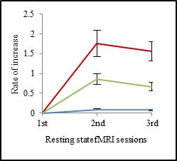
Animal Preparations: A total of 11 Sprague-Dawley rats (8-9 wks, male, 300 ± 20 g) were used in this study. Rats were anesthetized using isoflurane and intubated with a 16 gage vascular catheter to control physiology state. For arterial blood gas analysis (ABGA), blood sampling performed on left femoral artery after every rs-fMRI acquisition. Rs-fMRI acquisition was performed in 3 sessions (Fig. 1): 1) immediately after setting the animal in the magnet, without any physiologic adjustment, uncontrolled condition; 2) after electric stimulated BOLD-fMRI acquisition was performed, normal physiologic condition (PCO2=35~45, PO2=80~100 and PH=7.35~7.45); 3) during abnormal physiologic condition, hypoventilated condition (i.e., hypercapnia, pCO2>45, pO2<80, and pH<7.35)). The 1st rs-fMRI acquisition was finished in about 30 minutes. For the 2nd rs-fMRI acquisition, in order to get normal and stable physiologic condition, we tried to acquire electric stimulated BOLD-fMRI data which needed about 3~6 hours to reach the normal and stable physiologic condition in the magnet. We confirmed the signal of electric forepaw stimulated BOLD-fMRI (pulse width= 1.0 ms, current= 1.4 mA) at normal physiologic condition. For the 3rd rs-fMRI acquisition, we reduced ventilation about 30% than normal level and increased anesthesia 1.2 to 2.0 %.
MRI data acquisitions: All MRI data were obtained using Bruker 7T animal scanner equipped with a quadrature birdcage coil and array brain coil. Rs- fMRI data were collected using a single-shot gradient echo EPI sequence (TE= 20 ms, TR= 1000 ms, flip angle= 45°, number of average= 1, field of view= 30 (readout) x 15 (phase encoding) mm2, matrix size= 64 x 64, in-plane resolution= 469 x 233 μm2, slice thickness= 1.0 mm, number of slices= 12 coronal slices, number of repetition= 380, scan time= 6 min 20 sec).
Data Analysis: All preprocessing were performed using the FMRIB Software Library (FSL) packages and Analysis of Functional NeuroImages (AFNI). The raw data were pre-treated with skull stripping, slice timing correction, motion alignment, linear registration, nuisance regression, band-pass filtering and spatial smoothing. We compared betweenness centrality (BC), degree centrality (DC) and eigenvector centrality (EC) by analyze voxel-wise functional networks correlation for each subject. We used a paired t-test to calculate statistical group differences in rs-fMRI sessions using SPSS 19.0 software.
Results and Discussion
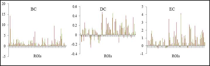
To evaluate resting state-fMRI functional connectivities, we calculated the growth rates in centralities for three rs-fMRI sessions. We measured BC, DC, and EC that indicate the number of the shortest path, the size of nodes, and correlation between nodes, respectively2. For the normal physiologic condition (2nd session)), higher for all the centralities (BC, DC, and EC) were observed compared to the 1st session (immediately after setting the animal in the magnet, without physiologic adjustment) (p < 0.05) and the 3rd session (hypoventilated condition) (p < 0.05) as shown in Figure 1. For the hypoventilated condition (3rd session), all the centralities (BC, DC, and EC) were lower than the 2nd session. (Figure 2)Conclusion
In most of rs-fMRI experiments in laboratories, adjustment of normal physiology for subjects before MRI scan is usually ignored by obtaining the data immediately after setting up the animal in the magnet. As indicated in this study, this may well result in insufficient rs-fMRI data acquisition, leading to lower functional connectivity data. Normal or abnormal physiologic conditions seem to have a direct effect on the rs-fMRI result. Thus, the results of our current study clearly suggest that adequate physiologic adjustment before rs-fMRI experiment is mandatory for rs-fMRI data acquisition.Acknowledgements
No acknowledgement found.References
1. Di X, Gohel S, Kim EH, et al. Task vs. rest-different network configurations between the coactivation and the resting-state brain networks. Front Hum Neurosci. 2013 Sep 17;7:493
2. Bullmore E, Sporns O., et al. Complex brain networks: graph theoretical analysis of structural and functional systems. Nat Rev Neurosci. 2009 Mar;10(3):86-98
Figures