5304
Effects of anesthesia on BOLD, electrophysiology, and PO2 signals in the whisker barrel cortexDaniil P Aksenov1, Limin Li1, Michael Miller1, Robert Linsenmeier2, and Alice M Wyrwicz1
1NorthShore University HealthSystem, Evanston, IL, United States, 2Northwestern University, Evanston, IL, United States
Synopsis
A comprehensive understanding of the relationship between neuronal activity and oxygen dynamics in the brain is vital for the accurate interpretation of fMRI results. This study investigates the relationship between changes in blood and tissue oxygen as well as neuronal activity in the awake and anesthetized states. We compare BOLD, PO2 and electrophysiological signals in the barrel cortex during whisker stimulation in awake and isoflurane-anesthetized rabbits. Our findings suggest that the relationship between BOLD, PO2 and electrophysiological responses is considerably different in the awake and anesthetized states.
Introduction
Blood oxygenation level dependent (BOLD) functional magnetic resonance imaging (fMRI) relies upon the changes in oxy- and deoxyhemoglobin in the local brain vessels. A comprehensive understanding of the relationship between neuronal activity and oxygen dynamics in the brain is vital for the accurate interpretation of fMRI results and for the application of fMRI techniques to more sophisticated and detailed neurophysiological questions. For this purpose, the use of electrodes to record oxygen tension (PO2) allows us to measure the oxygenation changes that underlie the BOLD signal more directly, as compared to derived measures such as CMRO2. Here we investigate how changes in BOLD, PO2 and electrophysiological signals are related in both the awake and anesthetized states We have performed fMRI, electrophysiological and PO2 recordings in whisker barrel cortex of the rabbit during whisker stimulation both in the awake state and under isoflurane anesthesia. These results will help us to build a more detailed picture of the relationship between neuronal activity, cerebral oxygen metabolism, and the BOLD signal.Methods
Dutch-Belted rabbits were chronically implanted with manipulators containing a single bundle of 4 microwire gold-silver electrodes aimed at the whisker barrel cortex. MR imaging experiments were performed on a 9.4T Bruker BioSpec imaging spectrometer. fMRI data were acquired from four consecutive 1mm-thick slices in the coronal plane using a single-shot, gradient-echo multi-slice EPI sequence with a repetition time (TR) of 2 s, an echo time (TE) of 11 ms, a 30mm×30mm field of view (FOV), and a matrix size of 80×80, corresponding to an in-plane resolution of 375 μm x 375 μm. The slices included the whisker cortex and whisker thalamus. Whisker vibration was delivered at amplitude of ±1.0 mm with a frequency of 50 Hz. Each experiment consisted of 10 trials (20 s baseline; 20 s whisker stimulation; and 40 s recovery). Activated voxels were detected in the averaged data by cross-correlation to a boxcar function at a statistical threshold corresponding to p<.001. Neuronal activity was recorded using Neuralynx system. The signals were amplified, band-pass filtered (300 Hz-3 kHz for SU and 1-150 Hz for local field potentials (LFP)), and digitized (32 kHz/channel). Data were analyzed after removal of blocks of gradient interference. To record PO2 changes an oxygen sensitive electrode was polarized to −0.7 V with respect to a silver-chloride reference electrode, and the current was measured with a Keithley model 614 electrometer. BOLD activation and either PO2 or electrophysiological data were recorded simultaneously.Results
As shown in Fig. 1, the temporal behavior of the BOLD fMRI and PO2 responses to whisker stimulation in the awake state were quite similar. The duration of the response was similar, and both the BOLD and PO2 responses decreased below the initial baseline level in the post-stimulus recovery period. Isoflurane anesthesia (0.5%) affected the temporal behavior of both signals. The duration of the BOLD response appeared to decrease, and the presence of an initial peak following the onset of stimulation was evident in the time course. The duration of the PO2 response remained similar to the awake state, but no post-stimulus undershoot with respect of the baseline was observed and the increase in signal magnitude was more gradual over the course of stimulation. The shape of electrophysiological responses (Fig. 2A,B) did not necessarily match the shape of BOLD and PO2 responses in the awake state. The duration of the electrophysiological responses was shorter than the duration of both the BOLD fMRI and PO2 responses, decreasing to baseline shortly after the end of stimulation. During isoflurane anesthesia the baseline and response of the electrophysiological signals decreased but the duration and shape of the responses did not visibly change.Discussion
Oxygen consumption increases in the brain in response to elevated neuronal activity, and the fMRI BOLD signal is closely related to the cerebral blood flow that supplies oxygen to areas of neuronal activation. Thus, a good correlation between oxygen, electrophysiological, and BOLD signals would be expected. Our results, obtained from simultaneous acquisition of BOLD fMRI and PO2 data, indicated that while BOLD and PO2 responses are similar in the awake state, differences emerge during anesthesia. Furthermore, the temporal behavior of the BOLD and PO2 responses is often inconsistent with electrophysiology. These findings suggest that under normal conditions tissue PO2 corresponds to the vascular dynamics of oxygen, as captured by BOLD fMRI. However, anesthesia disrupts the relationship among neuronal activity, vascular and tissue oxygen levels. Thus, the balance of oxygen delivery, consumption, and neuronal activity does not appear to be maintained under isoflurane anesthesia.Acknowledgements
Supported by: R01GM112715, McCormick School of Engineering and Applied Science.References
No reference found.Figures
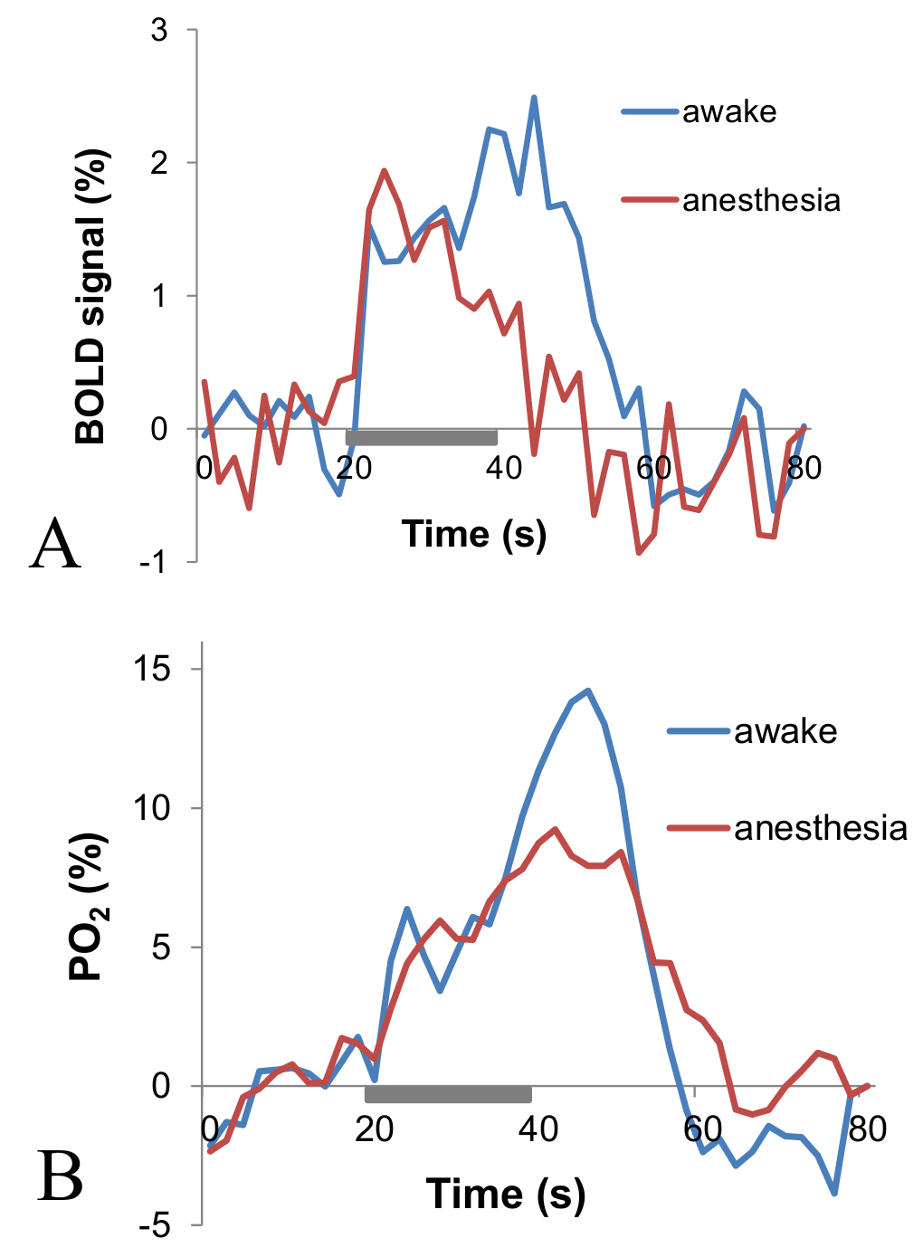
Fig. 1 Example of BOLD fMRI (A) and PO2
responses (B) to whisker stimulation in the whisker barrel cortex from a representative
animal in awake and anesthetized states.
The gray bar indicates the timing of the stimulus presentation.
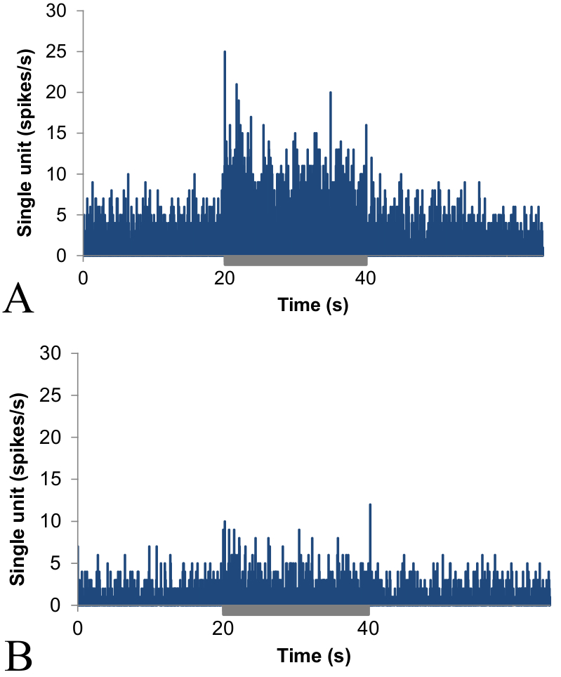
Fig. 2. Single unit responses to
whisker stimulation in the whisker barrel cortex in awake (A) and anesthetized
(B) states. SU baseline, peak, and plateau all decreased with anesthesia. The
gray bar indicates the timing of the stimulus presentation.