5302
Retrospective Independent Component Estimation of Respiratory and Cardiac Artefact Residuals (RICERCAR) in BOLD-fMRI.1UCL, London, United Kingdom
Synopsis
Variations in the heart and respiration rate have an impact on BOLD-fMRI signal variations. The cardiac cycle causes a pulsatile arterial blood flow which causes slice-specific signal changes resulting in artificial correlations between voxels within the same slice. The introduction of multi-band (MB) EPI acquisitions such as in the Human Connectome Project (HCP) increase such artificial correlations because many slices are acquired at the same slice time. We find physiological-related spatial Independent Components (ICs) and remove their corresponding time courses from BOLD-fMRI scans. Our method RICERCAR outperforms RETROICOR as well as FIX.
PURPOSE
Multi-band (MB) echo-planar imaging (EPI) provides an increased acquisition frequency for BOLD-fMRI resulting in a better temporal signal-to-noise ratio (tSNR) and better identification of details of the hemodynamic response (HRF) such as the early dip. However, EPI has the shortcoming that individual slices are differently influenced by the cardiac and respiratory cycle, which potentially results in artificial correlation between voxels in one slice. Such artificial correlations are multiplied in MB because multiple slices are acquired at the same slice timing. We perform spatial ICA and associate each IC to the power of cardiac or respiratory frequencies in the slice-wise global signal (SWGS), and show that removal of relevant physiology ICs result in diminished physiological frequencies in the scan.METHODS
We compute the SWGS to obtain information about the power of cardiac and respiratory frequencies in BOLD-fMRI. We average the signal of all voxels within a brain mask acquired with the same slice time and then compute the time-frequency power spectrum applying a short-window Fourier Transform. We obtain cardiac and respiratory frequencies from physiological recordings. We estimate 40 spatial ICs with the FastICA algorithm [1]. The power change of cardiac or respiratory frequencies after linear regression of one IC’s time course specifies the relatedness of an IC to physiological noise. We take the 5 most cardiac-related and the 5 most respiratory-related ICs and remove them from the scan. We compare our proposed method RICERCAR to RETROICOR [2], HCP minimal preprocessing [3] in MNI and FIX [4,5]. Our RETROICOR model comprises the low-frequency Fourier series expansion up to order to 2 of physiological recordings of cardiac and respiratory cycle [2]. Additionally, We convolve respiratory volume (RV) with the respiratory response function (RRF) and convolve the heart rate (HR) with the cardiac response function (CRF) to obtain 2 additional regressors [6,7]. We do not compute the FIX model but investigate HCP minimal-preprocessed and FIX-preprocessed data provided by the HCP. FIX is a data-driven approach applied after BOLD-fMRI scans were preprocessed with HCP minimal. We perform our own non-linear registration to map the slice timings from the original imaging space to the MNI space with nearest neighbor interpolation. This allows us to compute the SWGS in MNI space and compare RICERCAR to HCP minimal and FIX preprocessing.RESULTS
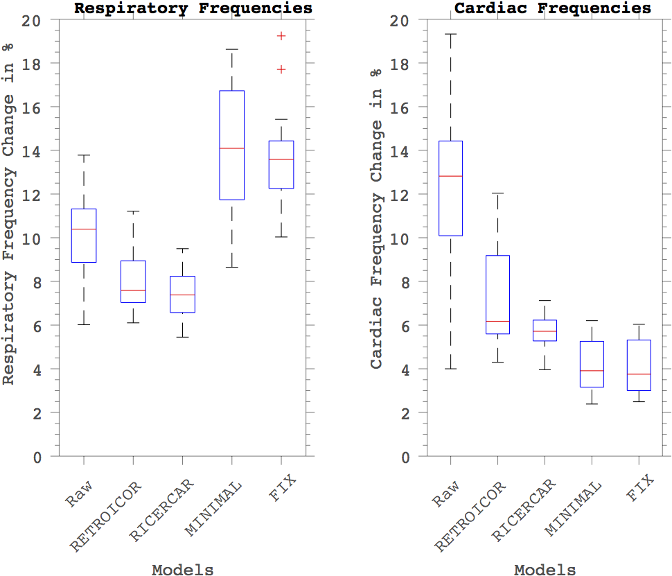
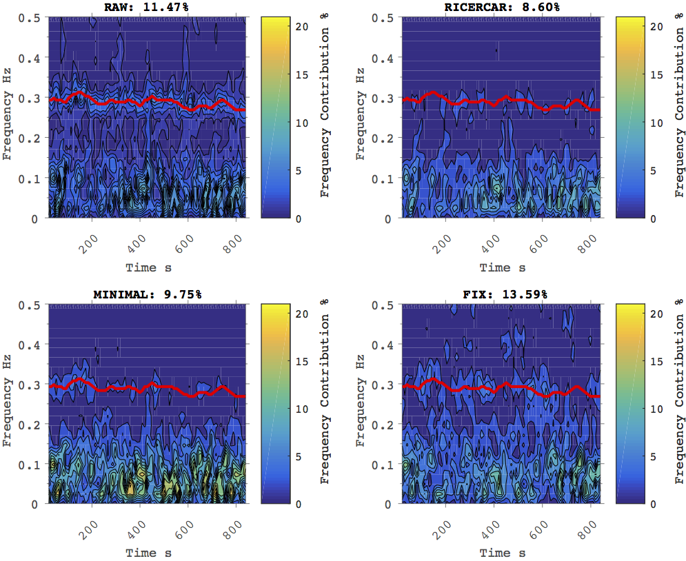
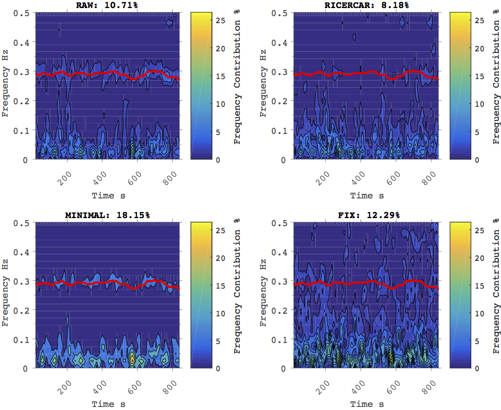
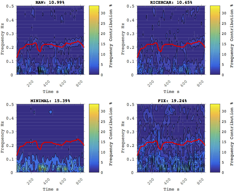
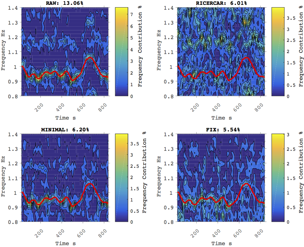
We examined 10 unrelated subjects from HCP comprising two BOLD-fMRI 15min scans per subject. Figure 1 summarizes the score of respiratory and cardiac frequency contributions before and after preprocessing with individual techniques. Cardiac and respiratory frequencies have a significant power contribution in the SWGS in all examined raw scans. Preprocessing with RETROICOR or RICERCAR achieves a great reduction in cardiac and respiratory related frequencies in the original image space as depicted in Figure 2,3,4,5 (top row). Scans preprocessed and mapped to MNI with the HCP minimal preprocessing and subsequent FIX artefact removal still contain physiological slice-artefacts that manifest in heart and respiration frequency contributions in the SWGS as depicted in Figure 2,3,4,5 (bottom row). Cardiac related frequencies are substantially lowered after mapping to MNI. However, the characteristic shape of the heart rate still remained in some scans seen in Figure 5 (bottom row).
DISCUSSIONS
The relative small reduction in physiological frequencies present in MNI space after FIX indicates that FIX is not very effective. The relative contribution of respiratory frequencies rises in MNI space whereas the contribution of cardiac frequencies diminishes in MNI. The increase in respiratory frequencies could be explained by the removal of other noise sources such as motion in the HCP minimal preprocessing pipeline, and therefore, results in an increase of respiratory frequencies in the SWGS. The reduction in cardiac frequencies is likely due to interpolation of adjacent slices acquired at different times. An unpredictable non-linear mixture of signal occurs and potentially results in structured physiological noise components comprising arteficial correlations among voxels acquired at the same time. Data-driven approaches such as FIX require physiological prior information in MNI space to work. In contrast, RICERCAR accounts for individual subject variability in physiology and creates a direct link between physiological recordings and ICs. Physiological slice artefacts can be removed in the original image space before further analysis.
CONCLUSIONS
We provide an efficient method to remove physiological slice-specific artefacts from HCP data without the requirement of non-linear registration into MNI space. We have shown that heart and respiration related frequencies are significantly removed in power, outperforming RETROICOR and FIX. Our method RICERCAR is thus a crucial contribution to preprocessing of individual subjects before studying their functional connectivity patterns in their original acquisition space.Acknowledgements
No acknowledgement found.References
[1] Beckmann, C.F. and Smith, S.M., 2004. Probabilistic independent component analysis for functional magnetic resonance imaging. IEEE transactions on medical imaging, 23(2), pp.137-152.
[2] Glover, G.H., Li, T.Q. and Ress, D., 2000. Image-based method for retrospective correction of physiological motion effects in fMRI: RETROICOR. Magnetic resonance in medicine, 44(1), pp.162-167.
[3] Glasser, M.F., Sotiropoulos, S.N., Wilson, J.A., Coalson, T.S., Fischl, B., Andersson, J.L., Xu, J., Jbabdi, S., Webster, M., Polimeni, J.R. and Van Essen, D.C., 2013. The minimal preprocessing pipelines for the Human Connectome Project. Neuroimage, 80, pp.105-124.
[4] Griffanti, L., Salimi-Khorshidi, G., Beckmann, C.F., Auerbach, E.J., Douaud, G., Sexton, C.E., Zsoldos, E., Ebmeier, K.P., Filippini, N., Mackay, C.E. and Moeller, S., 2014. ICA-based artefact removal and accelerated fMRI acquisition for improved resting state network imaging. Neuroimage, 95, pp.232-247.
[5] Salimi-Khorshidi, G., Douaud, G., Beckmann, C.F., Glasser, M.F., Griffanti, L. and Smith, S.M., 2014. Automatic denoising of functional MRI data: combining independent component analysis and hierarchical fusion of classifiers. Neuroimage, 90, pp.449-468.
[6] Birn, R.M., Smith, M.A., Jones, T.B. and Bandettini, P.A., 2008. The respiration response function: the temporal dynamics of fMRI signal fluctuations related to changes in respiration. Neuroimage, 40(2), pp.644-654.
[7] Chang, C., Cunningham, J.P. and Glover, G.H., 2009. Influence of heart rate on the BOLD signal: the cardiac response function. Neuroimage, 44(3), pp.857-869.
Figures