5287
Oxytocin influences the directed connectivity between the precuneus and the dorsolateral prefrontal cortex1Division of Psychiatry and Applied Psychology, University of Nottingham, Nottingham, United Kingdom, 2Radiological Sciences, Division of Clinical Neuroscience, University of Nottingham, Nottingham, United Kingdom, 3Department of Psychiatry & Robarts Research Institute, University of Western Ontario, London, ON, Canada
Synopsis
The default mode network (DMN) is now known to play an important role in social cognition. Thus, we hypothesized that the social neuropeptide oxytocin (OXT) will modulate the connectivity of the DMN. We used Granger Causality Analysis and found that intranasal OXT modulates the effective or ‘causal’ connectivity between the precuneus: a key DMN node and the dorsolateral prefrontal cortex, a key region in the central executive network. Thus, OXT has the potential to enhance the cooperative role of the DMN, which could explain the mechanistic action by which OXT improves social cognition in disorders such as autism and schizophrenia.
Objective
Recent research has shown the involvement of the neuropeptide oxytocin (OXT) in social cognition and its effects on the social brain.1 The default-mode network (DMN), initially considered to be a task negative network, is now known to play an important role in the processing of socio-affective stimuli.2 Additionally, studies have shown that the DMN can cooperate with key networks such as the fronto-parietal central executive network (CEN) during the performance of certain cognitive tasks.3,4 In light of these findings, examining the effects of OXT on the connectivity of the DMN could prove to be beneficial in understanding how OXT modulates social cognition. Evidence from functional connectivity (Fc) studies show that intranasally administered OXT can affect the connectivity of the DMN,5,6 however the mechanistic process by which it modulates DMN function is not yet fully understood as Fc-based methods are limited in their capacity to study directions of relationship ("causal" or effective connectivity) between two brain regions. Hence, we used Granger Causality Analysis (GCA) to explore the effective connectivity of the precuneus: a central DMN node under the influence of intranasal OXT.Aim
Based on research that elucidates the cooperative role of the DMN, we aimed to investigate if OXT will modulate causal interactions between the precuneus and important areas of the CEN.Methods
15 healthy male volunteers (age range: 18-33 years) participated in this study. Using a randomized, double-blinded, placebo-controlled, crossover design, participants received 24 IU intranasal OXT or placebo (PLA) prior to a 5 minute resting-state functional MRI scan. All scans were acquired at Sir Peter Mansfield Imaging Centre, University of Nottingham using a Philips Achieva 3T MRI scanner. Resting-state data were pre-processed using DPARSFA7 using a standard pre-processing pipeline. Atlas-based precuneus masks were used as seed regions to perform bivariate first-order coefficient-based voxel-wise GCA analysis using the REST software.8 SPM8 was used to conduct paired t-tests on the GCA maps with an uncorrected threshold of p<0.05. Mean GCA coefficients were extracted for each participant by placing a 6mm sphere around the peak with the highest t value from these analyses (left dlPFC (ldlPFC) for both left and right precuneus seeds) and subject to one-sample t-tests in order to determine the direction of causal influence (positive or negative) for each condition - OXT and PLA. This 6mm sphere was then used as a secondary seed region and entered into another GCA in order to investigate the return causal influence to the bilateral precuneus from the ldlPFC. We also ran a bivariate Pearson correlation to investigate whether there is reciprocity in effective connectivity change between the bilateral precuneus and the ldlPFC as an effect of OXT influence. All statistical analyses were performed using SPSS 21 and used a bootstrapped (10,000 simulations) level of p<0.05.Results
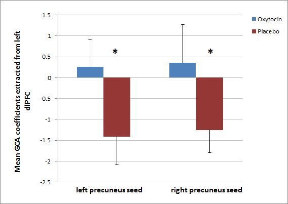
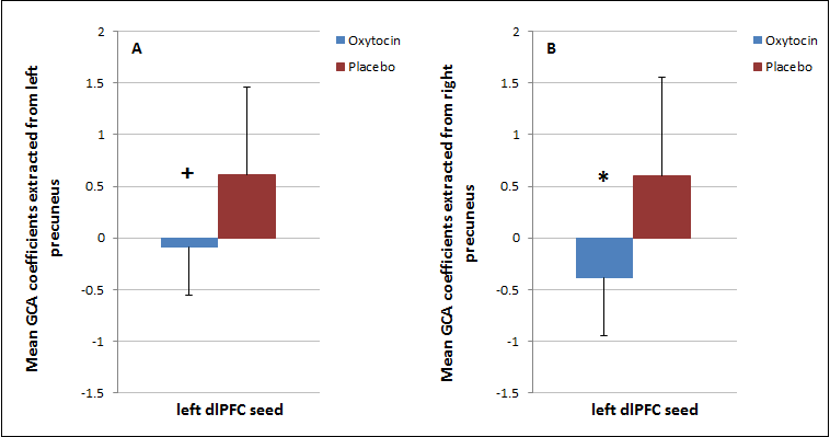
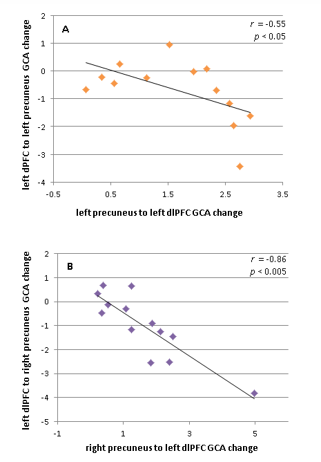
Voxelwise tests: OXT modulated the ‘causal’ outflow from both left and right precuneus to the ldlPFC (peak MNI co-ordinates: -39 9 45) (FWE cluster level p<0.005).Statistics performed on extracted mean GCA coefficients: One sample t-tests showed that under PLA, both the left and right precuneus exerted a significant negative causal influence on the ldlPFC (p<0.005). However, under OXT the bilateral precuneus exerted no significant causal influences on the ldlPFC (Figure 1). Paired t-tests revealed that OXT also significantly modulated the return ‘causal’ inflow from the ldlPFC to the right precuneus (p<0.05) and showed a trend for significance in modulating the inflow from the ldlPFC to the left precuneus (p=0.062). We found that under PLA, the ldlPFC exerted a significant positive causal influence back to the bilateral precuneus (p<0.05). However, under OXT, the ldlPFC exerted no significant causal influences on the left precuneus but exerted a significant negative causal influence on the right precuneus (p<0.05) (Figure 2). Additionally, we found a significant negative correlation between OXT induced changes in effective connectivity from precuneus-to-ldlPFC and ldlPFC-to-precuneus (left precuneus: r=-0.55, p<0.05; right precuneus: r=-0.86, p<0.005) i.e. reduction in negative influence (increase toward positive influence) from precuneus-ldlPFC was associated with a reduction in positive influence (increase toward negative flow) from ldlPFC-precuneus (Figure 3).
Discussion
A novel finding reported here is the effect of intranasally administered OXT on the directed influences between the precuneus - a core DMN node, and the dlPFC – a key central executive network region at rest. Thus, OXT has the potential to enhance the cooperative role of the DMN, which could explain the mechanistic action by which OXT improves social cognition in disorders such as autism and schizophrenia. These results encourage further research to unravel how OXT modulates effective connectivity in the brain during the performance of social cognition tasks.Acknowledgements
This study was supported by an Early Career Research Knowledge and Transfer Award from the University of Nottingham to Dr Birgit Völlm. We would like to acknowledge the assistance provided by Drs Mehri Kaviani and Elizabeth Liddle and Professor Peter Liddle in setting up this study and acquiring data.References
1. Zink CF, Meyer-Lindenberg A. Human neuroimaging of oxytocin and vasopressin in social cognition. Horm Behav. 2012; 61(3): 400-9. 2. Mars RB, et al. On the relationship between the "default mode network" and the "social brain". Front Hum Neurosci. 2012; 6:189. 3. Spreng RN, et al. Goal-congruent default network activity facilitates cognitive control. J Neurosci. 2014; 34(42): 14108-14. 4. Beaty RE, et al. Default and Executive Network Coupling Supports Creative Idea Production. Sci Rep. 2015; 5:10964. 5. Riem MM, et al. Oxytocin effects on complex brain networks are moderated by experiences of maternal love withdrawal. Eur Neuropsychopharmacol. 2013; 23(10): 1288-95. 6. Sripada CS, et al. Oxytocin enhances resting-state connectivity between amygdala and medial frontal cortex. Int J Neuropsychopharmacol. 2013; 16(2): 255-60. 7. Chao-Gan Y, et al, Yu-Feng Z. DPARSF: A MATLAB Toolbox for "Pipeline" Data Analysis of Resting-State fMRI. Front Syst Neurosci. 2010; 14, 4:13. 8. Song XW, et al. REST: a toolkit for resting-state functional magnetic resonance imaging data processing. PLoS One. 2011; 6(9):e25031.Figures