5283
Abnormal Heschl’s gyrus resting-state functional connectivity in patients with presbycusis1Shandong Medical Imaging Research Institute, Shandong University, Jinan, People's Republic of China, 2Philips Healthcare, Shanghai, People's Republic of China
Synopsis
This study revealed abnormal resting-state functional connectivity between the Heschl’s gyrus and distributed regions located in the hearing-related and language-related areas in patients with presbycusis. Our findings could be important for exploring imaging evidence of central component of presbycusis.
Purpose
Presbycusis is most often characterized by a progressive hearing loss at high frequencies, which are particularly important for speech recognition. Poor speech discrimination reflects a possible central component of presbycusis. Increased Heschl’s gyrus (HG) activities in patients with presbycusis in response to acoustical stimuli has been reported.1 However, there have been no prior studies using resting-state functional magnetic resonance imaging to investigate the HG functional connectivity (FC) in patients with presbycusis.Material and Methods
Fifteen patients with mild presbycusis (5 males/10 females, mean age, 63.2 ± 2.6 years) and fourteen age- and sex-matched healthy controls (6 males/8 females, mean age, 62.4 ± 2.0 years) were recruited in this study. Pure tone audiometry and tympanometry were used to assess the hearing abilities of all subjects. Hearing loss was defined as a speech-frequency pure-tone average (PTA) of thresholds at 0.5, 1, 2, and 4 kHz (air conduction) in the better-hearing ear. All subjects were scanned on a 3T scanner (Philips “Achieva” TX, Best, The Netherlands) and the rs-fMRI data were preprocessed. FC analysis was performed by using REST. A seed-to-voxel connectivity analysis was conducted which computed the correlation between seeds and other voxels. The seeds of bilateral HG were defined according to the automated anatomical labeling (AAL) template. Statistic analyses were performed using SPM8 by the application of multiple comparisons and the setting of a threshold of p < 0.001 (uncorrected) at voxel level and a threshold of p < 0.05 (FWE corrected) at cluster-level.Results
All patients had a type-A curve on tympanometry, which indicated normal middle-ear function. The PTA was significantly higher in patients with presbycusis than in healthy patients (p < 0.001). In the normal control group, the mean hearing thresholds were less than 20 dB HL at all frequencies; in the presbycusis group, the mean hearing thresholds were more than 20 dB HL at 1 kHz and reached 46.8 dB HL at 4 kHz and 55.2 dB HL at 8 kHz (Fig. 1). Patients with presbycusis had a lower left HG functional connectivity with the left superior temporal gyrus (Wernicke's area), middle temporal gyrus, temporal pole, insula and supramarginal gyrus (Wernicke's area) and a higher left HG functional connectivity with the left superior and middle frontal gyrus relative to healthy controls. Patients with presbycusis had a lower right HG functional connectivity with the right superior temporal gyrus, middle temporal gyrus and insula relative to healthy controls (Fig. 2).Discussion
In this study, patients with presbycusis had lower FC between the left HG and two language-related areas within the left superior temporal gyrus and supramarginal gyrus. This finding is consistent with previous studies: decreased audibility in patients with presbycusis influences speech processing, especially in noisy environments.2 The FC between the left and right HG and insula were also lower in patients with presbycusis compared with control subjects. This is consistent with a number of functional imaging studies that have shown that the insula plays an important role in auditory processing.3 The higher FC between the left HG and frontal lobes of patients with presbycusis may be a mechanism that helps to compensate for the lost ability to inhibit background noise.4Conclusion
This study revealed abnormal FC between the HG and distributed regions located in the hearing-related and language-related areas in patients with presbycusis. Our findings could be important for exploring imaging evidence of central component of presbycusis.Acknowledgements
This project was supported by the National Natural Science Foundation of China (grant nos. 81601479) and Shandong Provincial Natural Science Foundation of China (grant nos. BS2015YY003) and Shandong Provincial Medical and Healthy Technology Development Program of China (grant nos. 2015WS0176).References
1. Profant O, Tintera J, Balogova Z, et al. Functional changes in the human auditory cortex in ageing. Plos One 2015;10:e0116692
2. Snell KB, Frisina DR. Relationships among age-related differences in gap detection and word recognition. J Acoust Soc Am 2000;107:1615-1626
3. Bamiou DE, Musiek FE, Luxon LM. The insula (Island of Reil) and its role in auditory processing. Literature review. Brain Res Brain Res Rev 2003;42:143-154
4. Ouda L, Profant O, Syka J. Age-related changes in the central auditory system. Cell and Tissue Research 2015;361:337-358
Figures
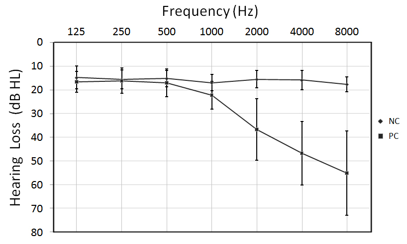
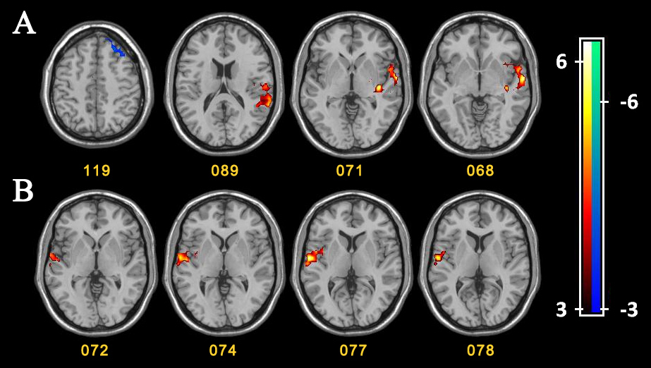
FIG 2. A. Functional connectivity (FC) analysis of the left Heschl’s gyrus (HG). Compared with healthy controls, patients with presbycusis had lower FC between the left HG and the left superior temporal gyrus, middle temporal gyrus, temporal pole, insula and supramarginal gyrus and higher FC between the left HG and the left superior and middle frontal gyrus.
B. FC analysis of the right HG. Compared with healthy controls, patients with presbycusis had lower FC between the right HG and the right superior temporal gyrus, middle temporal gyrus and insula.