5280
Tinnitus distress is linked to enhanced resting-state functional connectivity from the limbic system to the auditory cortex1Department of Radiology, Nanjing First Hospital, Nanjing Medical University, Nanjing, China, Nanjing, People's Republic of China, 2Nanjing First Hospital, Nanjing Medical University, Nanjing, China, Nanjing, People's Republic of China
Synopsis
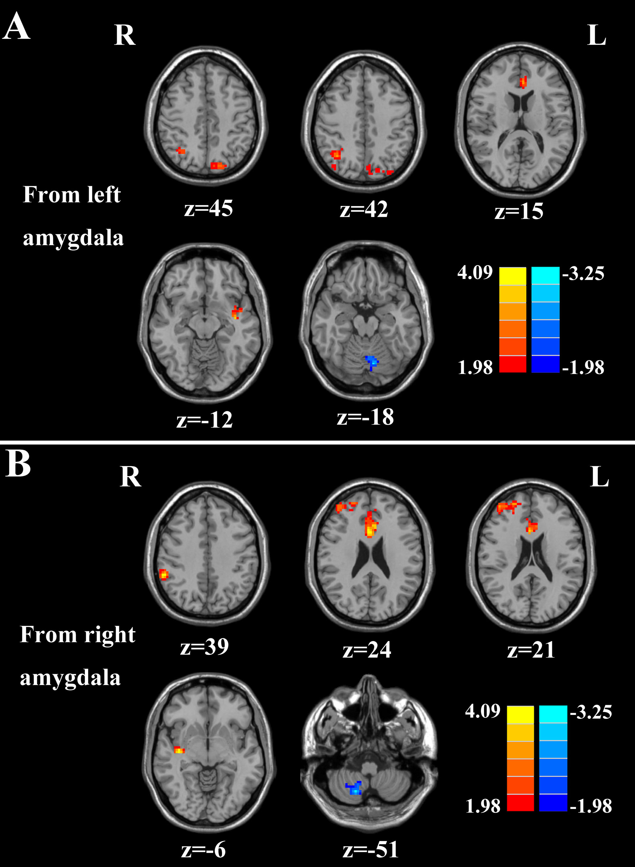
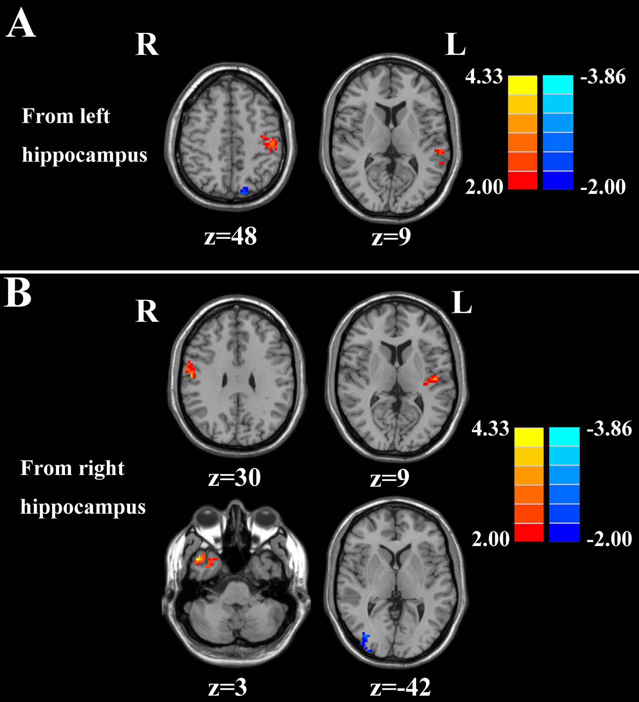
Our results identified disrupted effective connectivity networks in the limbic regions of tinnitus patients. Tinnitus severity was positively correlated with a bilateral increase in effective connectivity from the amygdala to the auditory cortex on the same side. In addition, tinnitus duration was positively correlated with enhanced effective connectivity from the right hippocampus to the left auditory cortex. These findings mainly emphasized the crucial role of limbic system and limbic-auditory interaction in tinnitus patients, which could help enhance our understanding of the neuropathological mechanisms underlying tinnitus.
Purpose
The phantom sound of tinnitus is believed to be triggered by aberrant neural activity in the central auditory pathway, but since this debilitating condition is often associated with emotional distress and anxiety, these comorbid symptoms likely arise from maladaptive functional connections to limbic structures such as the amygdala and hippocampus. To test this hypothesis, we used resting-state functional magnetic resonance imaging (fMRI) to identify aberrant effective connectivity between the amygdala and hippocampus and other brain regions in chronic tinnitus patients and determined the relationship of these abnormalities with specific tinnitus characteristics.Methods
Chronic tinnitus patients (n=26) and age-, sex-, and education-matched healthy controls (n=23) were included in this study. Both groups had clinically normal hearing thresholds. We used Granger causality analysis (GCA) with seed regions in the amygdala and hippocampus to identify abnormal brain effective connectivity in patients. The aberrant effective connectivity measures were then compared to other tinnitus metrics such as duration or distress.Results
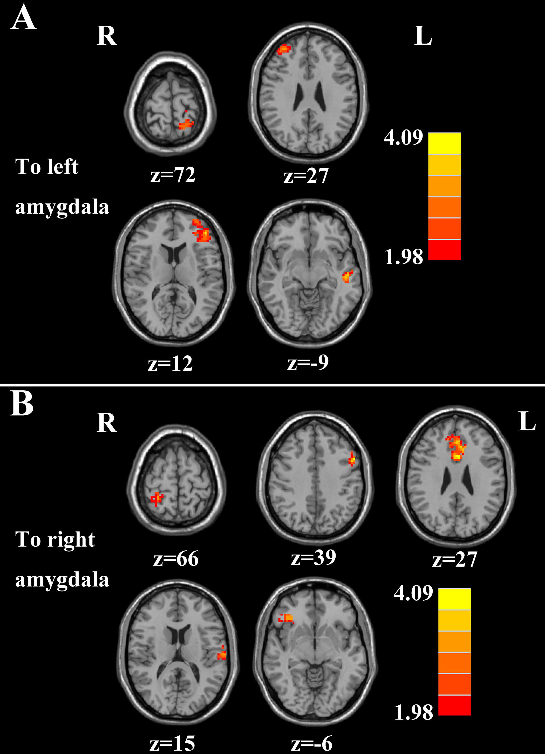
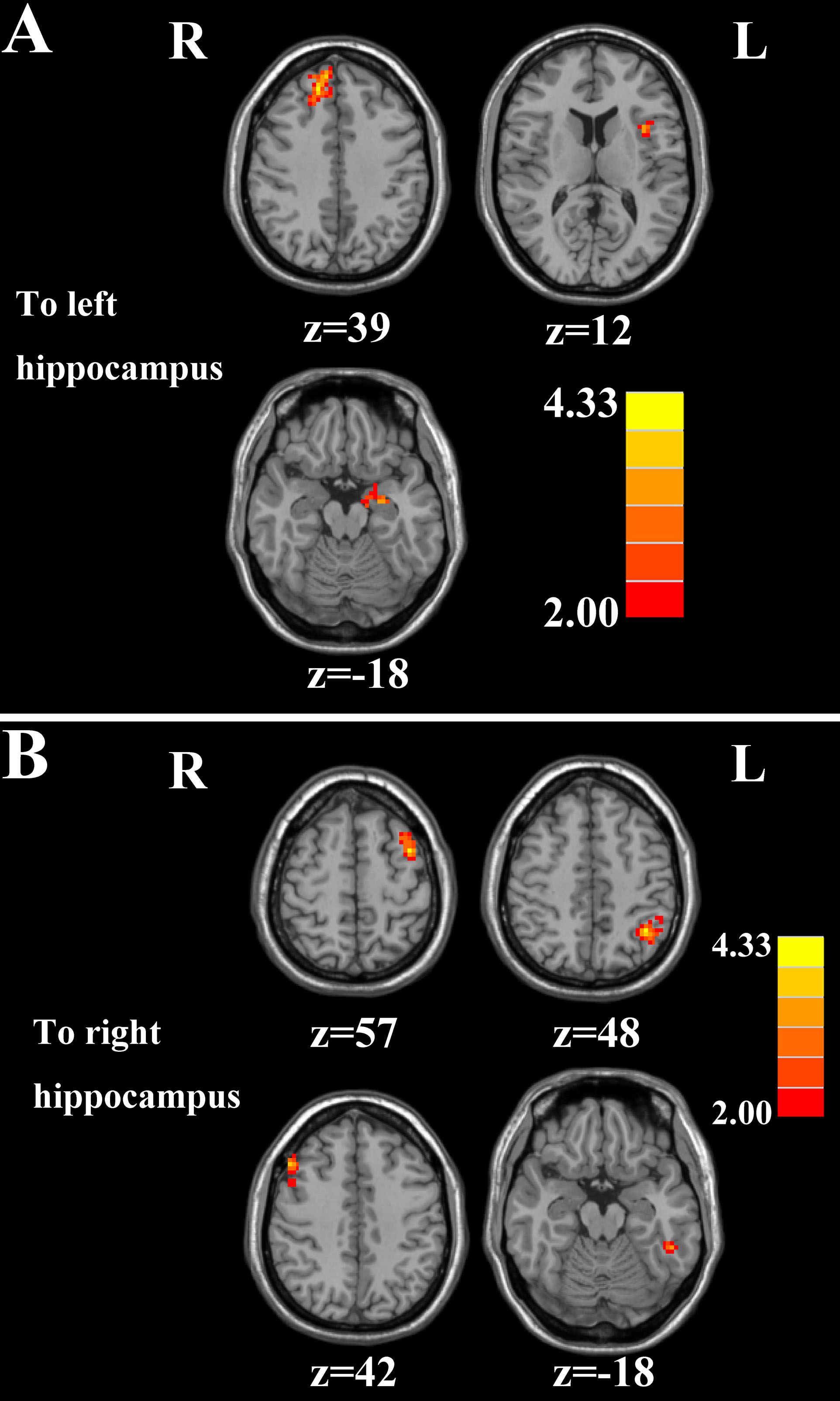
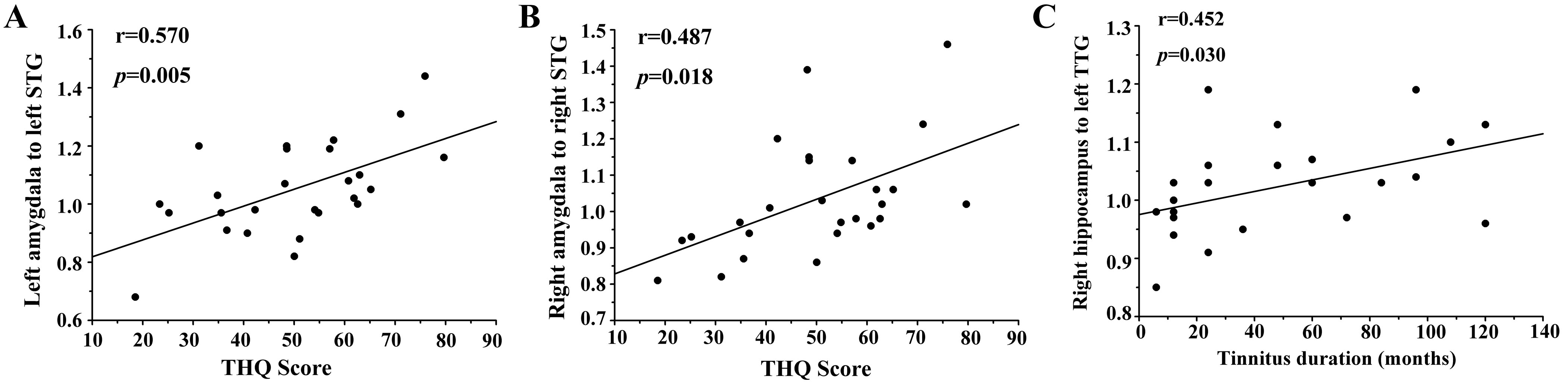
Relative to healthy controls, tinnitus patients demonstrated abnormal directional connectivity both from and to the amygdala and hippocampus; the main abnormalities were associated with primary and association auditory cortex, frontal cortex, anterior cingulate gyrus, and several other non-auditory areas. Importantly, scores on the Tinnitus Handicap Questionnaires were positively correlated with increased effective connectivity from the left amygdala to the left superior temporal gyrus (r=0.570, p=0.005), and from the right amygdala to the right superior temporal gyrus (r=0.487, p=0.018). In addition, enhanced effective connectivity from the right hippocampus to the left transverse temporal gyrus, an auditory area, was positively correlated with tinnitus duration (r=0.452, p=0.030).Discussions
To our knowledge, this is the first study to use GCA to identify changes in the direction of effective connectivity in two regions strongly implicated in tinnitus, the hippocampus and amygdala. In tinnitus patients, significant changes in effective connectivity occurred in a relatively circumscribed network emanating from and projecting to the amygdala or the hippocampus. In most cases, tinnitus was associated with an increase in effective connectivity rather than a decrease. A major finding of this study was that tinnitus distress was strongly correlated with a bilateral increase in effective connectivity from the amygdala to the STG. This suggests that the amygdala sends a strong negative emotional signal to the auditory cortex which influences how acoustic information is interpreted (LeDoux, 2007). The second major finding was that tinnitus duration was strongly correlated with increased effective connectivity from the right hippocampus to the left TTG. One interpretation of this result is that the hippocampus relays the memory of the phantom sound to the auditory cortex where acoustic image is consolidated to a chronic state (Halford and Anderson, 1991, Kraus and Canlon, 2012).Conclusions
Our results show that tinnitus distress is strongly correlated with enhanced effective connectivity that is directed from the amygdala to the primary auditory cortex. The longer the phantom sensation, the more likely acute tinnitus becomes permanently encoded (chronic) by memory traces in the hippocampus. These findings mainly emphasized the crucial role of limbic system and limbic-auditory interaction in tinnitus patients, which could help enhance our understanding of the neuropathological mechanisms underlying tinnitus.Acknowledgements
This work was supported by a grant from the National Natural Science Foundation of China (Nos. 81601477, 81600638) and Natural Science Foundation of Jiangsu Higher Education Institutions (No. 16KJB320001).References
LeDoux J (2007) The amygdala. Current Biology 17:R868-R874.
Halford JB, Anderson SD (1991) Anxiety and depression in tinnitus sufferers. Journal of psychosomatic research 35:383-390.
Kraus KS, Canlon B (2012) Neuronal connectivity and interactions between the auditory and limbic systems. Effects of noise and tinnitus. Hearing research 288:34-46.
Figures