5279
Increased resting-state functional connectivity within subgenual anterior cingulate cortex network reveals the neural substrate of emotion regulation following mindfulness-based stress reduction (MBSR) training1Institute of Biomedical Engineering, National Taiwan University, Taipei, Taiwan, 2Institute of Medical Device and Imaging, National Taiwan University College of Medicine, Taipei, Taiwan, 3Graduate Institute of Brain and Mind Sciences, National Taiwan University College of Medicine, Taipei, Taiwan, 4Department of Neurology, Cardinal Tien Hospital Yonghe Branch, New Taipei City, Taiwan, 5Department of Buddhist Studies, Dharma Drum Institute of Liberal Arts, New Taipei City, Taiwan, 6Department of Mass Communication, Tamkang University, Taipei, Taiwan, 7Department of Electrical Engineering, National Taiwan University, Taipei, Taiwan, 8Molecular Imaging Center, National Taiwan University, Taipei, Taiwan
Synopsis
Mindfulness-based stress reduction (MBSR) has been known to improve emotion regulation and cure mood disorders. The present longitudinal study aimed to clarify the effects of MBSR training and practice on the neural substrates of emotion regulation. Resting-state functional MRI (rsfMRI) exams were performed at three time points to investigate changes of functional connectivity (FC) within the subgenual anterior cingulate cortex (sgACC) network. MBSR training and practice strengthened the FC of sgACC with the inferior frontal gyrus and with the insula, and reduced the level of anxiety and depression. The findings reveal the neural substrates of emotion regulation process following MBSR.
PURPOSE:
Mindfulness-based stress reduction (MBSR) is modified from the Buddhist traditions and has been known to improve an individual’s emotion regulation and cure mood disorders1. However, the effect of MBSR training and practice on the neural substrate of emotion regulation is still unclear. To clarify this effect, we employed the resting-state functional MRI (rsfMRI) to investigate changes of functional connectivity (FC) within the subgenual anterior cingulate cortex (sgACC) network, which plays an important role in regulating emotion2. In the present study, all subjects received MRI exams at three different time points: one before the 8-week MBSR training, one after the training, and one at two months after the 2nd exam. During the period between the 2nd and 3rd exams, all subjects stopped any MBSR training and practice. Each MRI exam consisted of T1W imaging and two runs of 6-minute rsfMRI. In the period between the two runs of rsfMRI, subjects were asked to perform MBSR practice (self-awareness of breath) for 20 minutes. We hypothesized that: 1) the strength of FC within the sgACC network would change after the 20-minute MBSR practice within each MRI exam; 2) the degree of FC change within the sgACC network due to the 20-minute MBSR practice would be significantly different among three MRI exams.Methods:
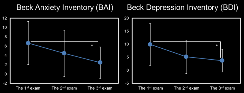
Study design: Eleven subjects (7 females and 4 males, age = 41.09±8.19 years), who were beginners of MBSR practice, participated the study and received an 8-week MBSR training. The detailed study design has been mentioned in the above section and shown in Figure 1. All subjects received the Beck Anxiety Inventory (BAI) and Beck Depression Inventory (BDI) tests for measuring the severity of anxiety and depression before each MRI exam. MRI data acquisition: MRI scanning was performed on a 3T MRI system (TIM Trio, Siemens) with a 32 channel phased-array head coil. T1W imaging was performed using a 3D-MPRAGE sequence: TR/TE = 2000 ms/3 ms, flip angle = 9 degree, FOV = 256×192×208 mm^3, matrix size = 256×192×208, and spatial resolution = 1 mm^3. Six-minute rsfMRI was performed using a 2D gradient EPI sequence: TR/TE = 2000 ms/24 ms, flip angle = 90 degree, FOV = 256×256×3 mm^3, matrix size = 64×64×34, and 180 volumes per run. Data processing: rsfMRI data was analyzed using data processing assistant for resting state fMRI (DPARSF) based on SPM8. Figure 2 shows the detailed procedure of data preprocessing and analysis. The seed-based analysis was performed to obtain whole-brain FC maps by placing a spherical seed in the bilateral sgACC3 (the center coordinate: [0, 25, -10], radius = 8 mm). Z-maps of sgACC FC were determined by computing Pearson correlation between the time course at each voxel of the brain and the averaged time course of the predefined sgACC seed, followed by the Fisher Z-transform. The change of sgACC FC Z-maps due to MBSR practice was computed for each MRI exam: ∆Z-map = Z-mappost-practice − Z-mappre-practice. We performed a non-parametric permutation ANOVA4 to detect brain regions with significant difference in ∆Z-maps among the three exams. Averaged ∆Z-values were then extracted from these brain regions to perform post-hoc tests using Mann-Whitney U-test.Results:
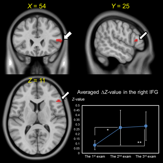
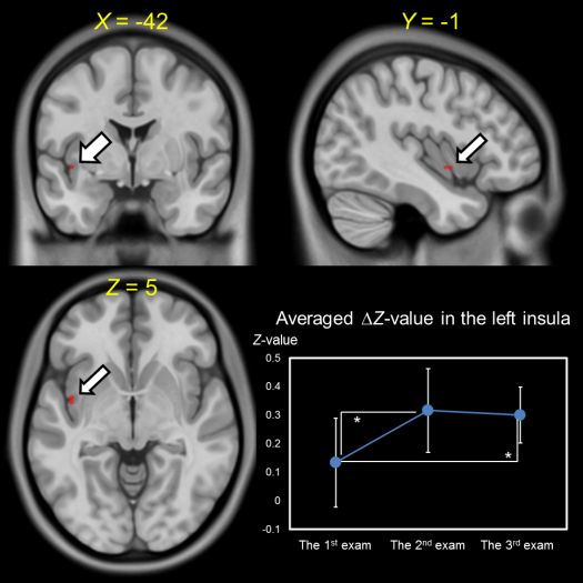
The right inferior frontal gyrus (IFG) and left insula showed significant difference in ∆Z-maps among three exams (non-parametric p < 0.05, uncorrected; Fig. 3 & 4). Compared with the 1st exam, the 2nd and 3rd exams showed significantly increased averaged ∆Z-values in both the right IFG and left insula. Moreover, compared with the 1st and 2nd exams, the results of both BAI and BDI showed significantly decreased scores in the 3rd exam (Fig. 5).Discussion:
We found enhancement of both the right sgACC-IFG FC and left sgACC-insula FC in the 2nd exam, and such enhancement still persisted in the 3rd exam. We also found persistent decrease in BAI and BDI scores following MBSR training. These findings imply a lasting effect of MBSR on brain connectivity and behaviors even after cessation of MBSR training. IFG is a component of the central executive network and relates to suppression of negative emotion5. Previous study suggests greater right sgACC-IFG FC might be better regulating negative emotional thoughts in depressed adolescents3. Both sgACC and insula are main components of the salience network and relate to integration of an individual’s emotional state and external stimuli6.Conclusion:
MBSR training and practice strengthen the sgACC-IFG FC and sgACC-insula FC, and reduce the level of anxiety and depression. The findings reveal the neural substrates of emotion regulation process following MBSR.Acknowledgements
No acknowledgement found.References
1. Grossman P, Niemann L, Schmidt S, et al. Mindfulness-based stress reduction and health benefits: A meta-analysis. J Psychosom Res. 2004;57(1):35-43.
2. Johansen-Berg H, Gutnab DA, Behrens TEJ, et al. Anatomical connectivity of the subgenual cingulate region targeted with deep brain stimulation for treatment-resistant depression. Cereb Cortex. 2008;(18):1374-1383.
3. Connolly CG, Wu J, Ho TC, et al. Resting-state functional connectivity subgenual anterior cingulate cortex in depressed adolescents. Biol Psychiatry. 2013; 74(12):898-907.
4. Nichols TE and Holmes AP. Nonparametric permutation tests for functional neuroimaging: a primer with examples. Hum Brain Mapp. 2002;(1):1-25.
5. Goldin PR, McRae K, Ramel W, et al. The neural bases of emotion regulation: Reappraisal and suppression of negative emotion. 2008;(63):577-586.
6. Uddin LQ. Salience processing and insular cortical function and dysfunction. Nat Rev Neurosci. 2015;(1):55-61.
Figures