5267
Evaluation of two novel MRI techniques for the assessment of intracranial pulsatility1Department of Engineering Physics, Tsinghua University, Beijing, People's Republic of China, 2Laboratory of FMRI Technology, Mark & Mary Stevens Neuroimaging and Informatics Institute,Keck School of Medicine,University of Southern California, University of Southern California, CA, United States, 3Key Laboratory of Particle and Radiation Imaging, Ministry of Education, Medical Physics and Engineering Institute, Department of Engineering Physics, Tsinghua University, Beijing, People's Republic of China
Synopsis
The brain pulsatility plays an important role in various cerebral pathology, such as brain tumor and traumatic brain injury. Two MRI methods have been recently developed for assessing brain biomechanical features using a ECG-gated cine sequence with different processing strategies. In this study, we evaluated and compared the two methods. Consistent findings were obtained using both methods that the majority of cardiac-induced brain pulsatile motion occurs in the brain stem and basal ganglia as well as in big arteries.
Purpose:
The alternation of intracranial pulsatility are related to a number of pathological changes, such as brain tumor and traumatic brain injury (TBI). Intracranial pulsatility can be assessed by the dynamic flow velocity curve of CSF using ECG-gated Phase-contrast MRI. Recently, two novel MRI methods have been proposed for the assessment of intracranial pulsatilities using a ECG-gated cine high-resolution acquisition: 1) Amplified MRI1 (aMRI), which uses Eulerian Video Magnification2 (EVM) to magnify the cardiac-included brain motion; and 2) Jacobian method3, which assesses the volumetric deformation across cardiac cycle using Jacobian determinant. The purpose of this study was to evaluate and compare the performance of the two MRI methods for the assessment of brain pulsatility across the cardiac cycle.Methods:
Data acquisition: An ECG-gated cine 3D integrated-SSFP (iSSFP) sequence was performed. Compared to balanced SSFP, a dephasing gradient following readout gradient was applied in iSSFP, resulting in 2p dephasing within a single voxel. iSSFP effectively removes banding artifacts while maintaining the unique bSSFP tissue contrast and the high SNR efficiency by averaging the bSSFP signal profile. A series of 3D high-resolution whole brain volumes across cardiac cycle was acquired using the ECG-gated cine 3D iSSFP sequences with the imaging parameters: FOV=220x165mm2, voxel size=1x1x2mm3, TR=4.8ms, TE=2.4ms, flip angle=35°, acceleration factor=2, 80 slices with slice thickness of 2mm. 6 to 9 brain volumes were collected over cardiac cycle with a temporal resolution of 105ms, within a total scan time of approximate 6 to 8 min. A total of 6 healthy volunteers were scanned on a Siemens Prisma 3T scanner. Data processing: (I) Amplified MRI: Eulerian video magnification technique2 (EVM) was used to magnify cardiac-induced brain motion from the ECG-gated MRI data treated as ‘video input’, which were spatially decomposed into different spatial frequency components, followed by a temporal bandpass filter to each spatial component to extract the motion at the passband frequencies of interest. A pass band of 0.01–0.03 Hz was selected according to the previous study1. To optimize the performance of EVM method on our data, EVM was executed with difference choices of the amplification parameter a and spatial frequency cutoff l. Finally, the amplified signal was added to the original signal, and the spatial components were summed to synthesize the output cine data. Subtle motions over the cardiac cycle were amplified, allowing us to visualize tissue deformations and other physiological variations. Normalized variance maps (normalized variance calculated over the cardiac phases) were produced to show amplified motion. (II) Jacobian method: The voxel-wise volumetric deformation in the brain was estimated by warping the series of brain volume across the cardiac cycle onto the last volume using Advanced Normalization Tools (ANTs). The warping parameters were transformed to Jacobian determinant (JD) (JD=1: no deformation; JD>1: expansion; JD<1: contraction). The deformation map was generated by calculating the difference between maximal and minimal Jacobian across the cardiac cycle at each voxel.Results and Discussion:
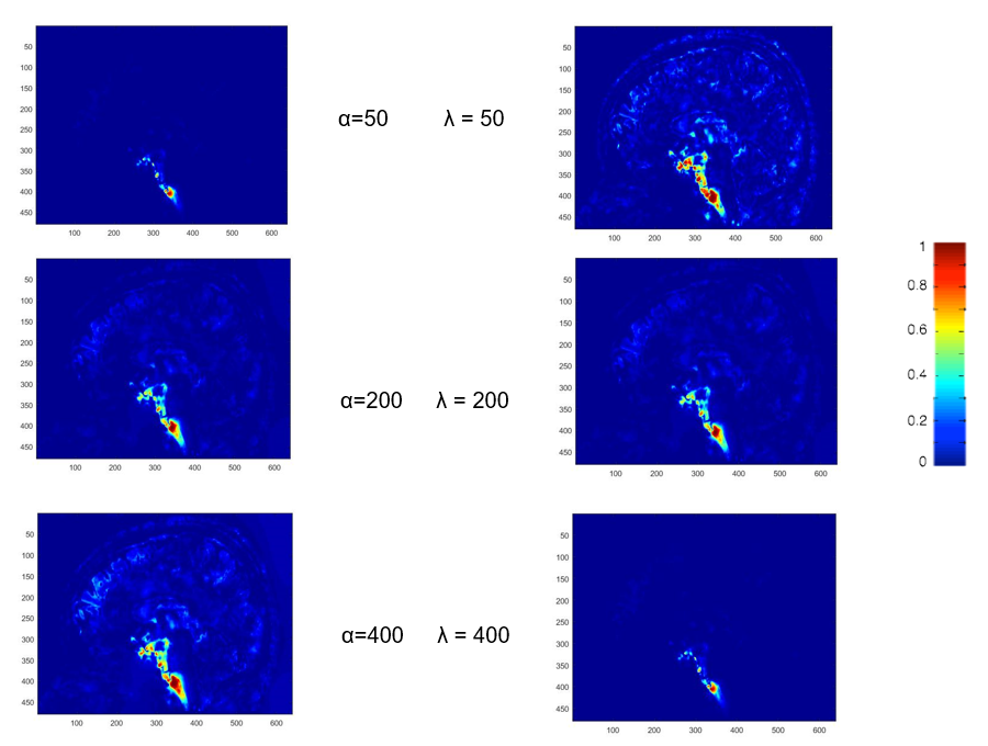
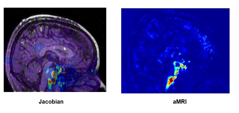
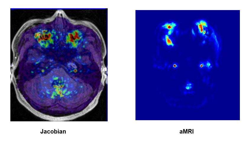
In amplified MRI, the proper selection of global amplification parameter α and spatial frequency cut-off wavelength λ is essential to achieve adequate motion amplification with low artifacts. Figure 1 shows the motion-amplified images with different sets of global amplification parameter α and spatial cutoff wavelength λ. With increasing α, the subtle brain motion becomes more apparent at the expense of increasing image noise. On the other hand, lower values of λ produce stronger amplification and higher noise. When λ is too high, motion of finer structures is not properly amplified. λ = 200 mm and α=200 were chosen for all the data processing. The cardiac-induced brain pulsation was assessed using both methods from all the subjects. Figure 2 shows the pulsatile motion at the same slice position using the two methods. Similar motion pattern was observed from the two methods. As one can appreciate, greater deformation occurs in the basal ganglia and brain stem compared the rest brain regions using both methods. Arterial pulsatility was also observed in big arteries, such as internal carotid arteries as shown in Figure 3. Our findings indicate the majority of brain pulsation occurs in basal ganglia and brain stem, which may result from the pulsatile arterial flow around circle of Willis and CSF bulk flow in ventricles.Conclusion:
Two recently introduced MRI methods for assessing intracranial pulsatility were evaluated in the present study. Both methods revealed that the majority of pulsatile motion occurs in basal ganglia and brain stem. The amplitude MRI and Jacobian methods may enable the characterization of brain biomechanical features in a wide range of brain diseases, such as brain tumor, TBI and Alzheimer’s disease.Acknowledgements
This work was supported by Tsinghua University Initiative Scientific Research Program.References
1. Samantha J. Holdsworth, Mahdi Salmani Rahimi, Wendy W. Ni, Greg Zaharchuk, and ichael E. Moseley. Amplified Magnetic Resonance Imaging. Magnetic Resonance in Medicine.2016;75:6.
2. Wu H, Rubinstein M, Shih E, Guttag J, Durand F, Freeman WT. Eulerian video magnification for 2revealing subtle changes in the world. ACM Trans Graph 2012;31:4.
3. Lirong Yan, Mayank Jog, Kay Jann, Xingfeng Shao, Danny JJ Wang. Brain pulsatility across the cardiac cycle revealed by cine 3D integrated-SSFP. ISMRM2016.
Figures