5245
Multiband Echo-SHift EPI (MESH-EPI): Applications at 3T1Donders Institute for Brain, Cognition and Behaviour, Radboud University Nijmegen, Nijmegen, Netherlands, 2Application Development, Siemens Healthcare, Erlangen, Germany, 3Faculty of Psychology and Neuroscience, Maastricht University, Maastricht, Netherlands, 4Maastricht Brain Imaging Center (MBIC), Maastricht University, Maastricht, Netherlands
Synopsis
In the current abstract we explore the applications of Multiband Echo-SHifted (MESH) EPI as a potential tool for fMRI at 3T. We compare MESH and SMS-EPI in two sets of experiments: the first looking into typical fMRI protocols, and the second with a focus in BOLD contrast and temporal resolution optimization. We conclude that MESH can increase the temporal efficiency of fMRI and be used to reduce distortions without sacrificing BOLD contrast.
Purpose
Demonstrate Multiband Echo Shift EPI, MESH-EPI[1], at 3T as a means to improve the temporal efficiency of fMRI acquisitions.Introduction
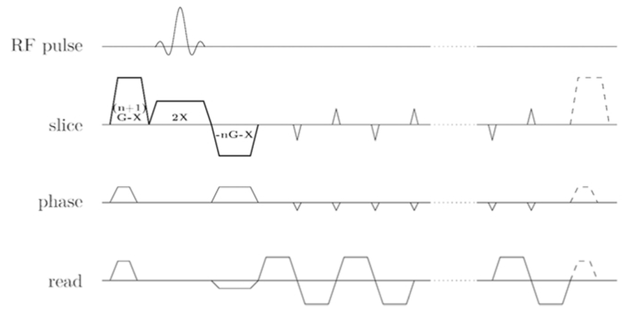
MESH is an interslice echo-shifted[2] simultaneous multi-slice (SMS)[3] with blipped-CAIPI[4] sequence developed to utilize the dead time that typically occurs in EPI fMRI acquisitions between the MB-RF pulse and the beginning of the readout train, necessary for the desired BOLD contrast to build up. In general, the TE should be as close as possible to the T_{2} star values of gray matter (over 50ms at 3T[5][6][7]). In traditional EPI acquisitions, extending the TE increases the TR, and thus penalizes the temporal resolution, and a short TE (e.g. 30ms) is often chosen as a compromise, and as a means to reduce through slice dephasing. The use of in-plane acceleration marginally improves the temporal resolution, and reduces image distortions, but increases the dead time. Unlike in-plane acceleration, SMS acceleration effectively reduces the TR, but by itself does not reduce distortions. MESH makes use of the extended dead time when in-plane acceleration and optimal TE are desired, and brings together (a) a BOLD optimized acquisition, (b) reduced image distortions, (c) compact sampling with echo-shifting and (d) TR reductions by the use of SMS acceleration. A diagram of the sequence, previously demonstrated at 1.5T [8] can be found in Figure 1.Methods
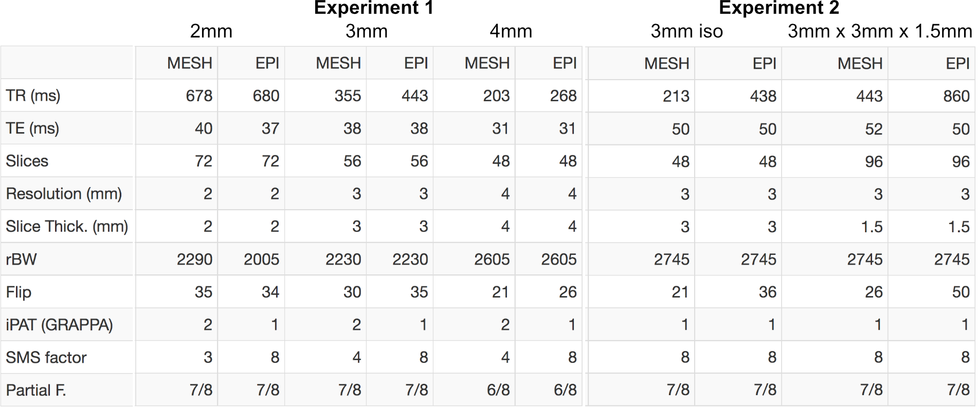
Firstly, we compared 3 pairs of protocols with different characteristics, close to the commonly found in SMS fMRI experiments. Each pair consisted of one standard SMS-EPI and one MESH-EPI protocols with similar parameters: a) 2mm isotropic resolutions with TRs close to those used by the Human Connectome Project[9]; b) 3mm isotropic resolutions allowing cardiac noise sampling; c) 4mm isotropic protocols with sampling times approaching those obtained by inverse imaging techniques[10].
Secondly, we compared 2 pairs of protocols with long TEs matching $$$T_{2}$$$ star values found in the literature and thus not requiring in-plane acceleration when using one echo-shift. Because long echo times are associated with signal dropouts, one pair of protocols was acquired with thin slices. Relevant parameters for the first and second experiments are listed in Figure2. All data were acquired from subjects after written consent on a MAGNETOM Prisma 3T MR scanner (Siemens Healthcare, Erlangen, Germany) using a 32-channel head-coil and a sequence derived from the standard product EPI. Images were reconstructed online using the SliceGRAPPA algorithm with LeakBlock[11].
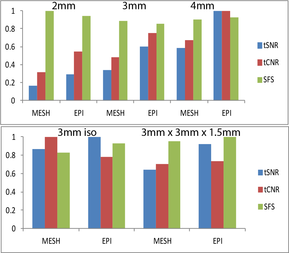
To validate our technique we computed the tSNR, the tCNR, defined as $$$tSNR * TE *\frac{e^{\frac{-TE}{T_{2}^{*}}}}{\sqrt{TR * vol}}$$$ and the Signal Fluctuation Sensitivity (SFS)[12] of all protocols. We expected a tSNR penalty for MESH because of either in-plane acceleration or the lower steady-state signal associated with lower flip angles. We also expected an increase in tCNR and ideally no loss in SFS in comparison to SMS-EPI. Both tSNR and tCNR values were averaged within a gray matter mask obtained from a T1 weighted structural scan.
Results
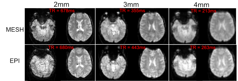
Images with contrast nearly identical to standard SMS-EPI, but with less distortions and an equal or better temporal resolution were obtained in the first set of experiments (see figure 2).
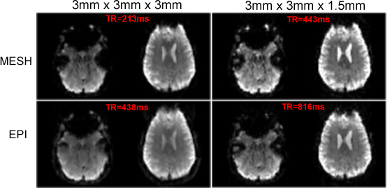
In our second experiment, with $$$TE=T_{2}^{*}$$$ of GM, MESH provided significant increases of temporal resolution (see figure 3).
Our quantitative evaluation (see figure 4) shows the expected drop in tSNR and tCNR, but no loss in SFS.
Discussion
In pair 3mm we showed fast sampling at the typical resolution of 3mm. The MESH protocol is fast enough to sample the cardiac rate (TR<380ms), and showed less distortions than a similar SMS-EPI protocol. For MESH images there is some degree of sensitivity reduction because of in-plane acceleration but at moderate acceleration factors this small compared to the physiological noise [13]. At 3mm isotropic with a 32ch head coil such losses can be virtually ignored[14]. In pair 2mm we matched the contrast and temporal resolution of a SMS-EPI protocol but reduced the distortions. In pair 4mm we pushed MESH to its limits to show an improvement in temporal resolution of about 30% in comparison to the equivalent SMS-EPI protocol. On our second experiment we showed that it is possible to extend the TE without sacrificing the temporal resolution or BOLD contrast. Contrary to our expectations, though, we did not see significant benefits when using thin slices.
MESH allows scans with less distortions and/or improved temporal resolution at 3T with virtually no sacrifice in SFS. To date we have only experienced using echo shifts of 1, higher shifts might allow bringing together those two benefits. Immediate applications include better separation of physiological noise for rsfMRI, temporal ICA and improved image registration.
Acknowledgements
The current work is supported by the European Union through a FP7-PEOPLE-2013-ITN grant as a part of a Marie-Curie "Initial Training Networks" Action.References
[1] Norris, D. 2013 [2] Ehses, P. 2014 [3] Larkman, D. 2001 [4] Setsompop, K. 2012 [5] Poser, B. 2006 [6] Peters, A. 2007 [7] van der Zwaag, W. 2009 [8] Boyacioglu, R. 2016 [9] van Essen, D. 2013 [10] Boyacioglu, R. 2013 [11] Cauley, S. 2014 [12] DeDora, D. 2016 [13] Zwart, J. 2002 [14] Triantafyllou, C. 2016
Figures