5235
Feasibility study of gradient echo recalled readout segmented EPI with VB-EPI for ultra-silent fMRI1University of Erlangen-Nuremberg, Erlangen, Germany, 2Siemens Healthcare GmbH, 3Fraunhofer MEVIS
Synopsis
We present an application of Variable-Blipped-EPI (VB-EPI) with readout-segmented EPI1, which has possible benefits for functional Magnetic Resonance Imaging (fMRI) experiments. Acoustic noise is reduced by lowering the amplitude of the readout (RO) gradient, which is possible due to the RO segmentation, and by prolonging the duration of the phase encoding (PE) blips simultaneously. Even with standard Cartesian parallel imaging techniques, like Generalized Autocalibrating Partially Parallel Acquisitions (GRAPPA)2, high resolution images can be obtained with high image quality.
Purpose:
In functional Magnetic Resonance Imaging (fMRI) typically Echo-Planar-Imaging (EPI)3 sequences are used to acquire whole volumes rapidly. This comes with a high level of acoustic noise, which affects fMRI experiments4. Acoustic noise can be reduced in EPI by using a sinusoidal readout (RO) gradient in combination with a constant phase-encoding (PE) gradient5. In our approach, instead of using a constant PE gradient we prolong the duration of the blips, called Variable-Blipped (VB)-EPI. This is combined with readout-segmented (rs) EPI, which can be used to further reduce acoustic noise by lowering the RO amplitude. With this combination it is possible to acquire high resolution EPI data with significantly reduced acoustic noise and high image quality.Method:
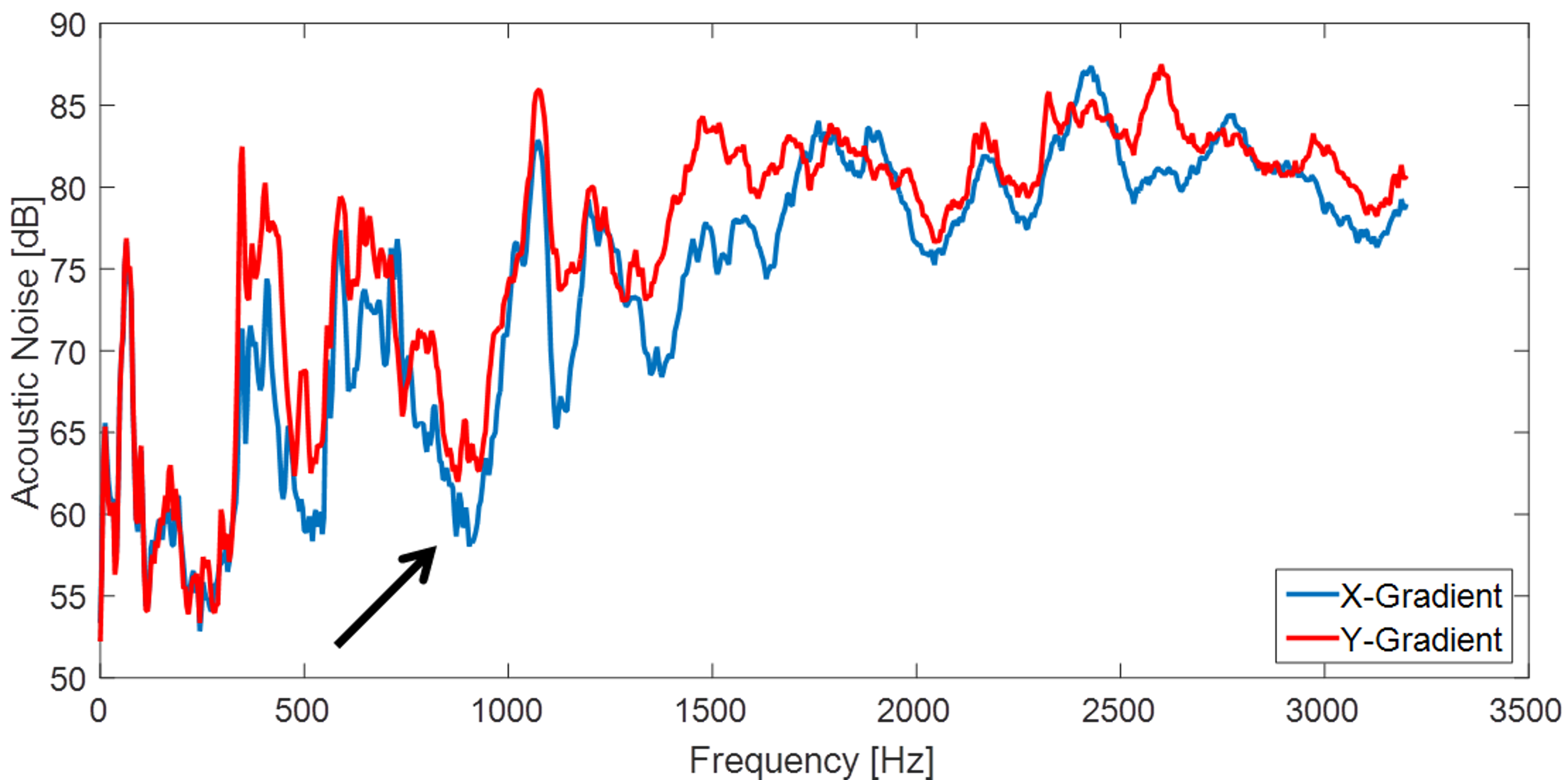
We use a prototype sequence based on a modified rs-EPI1 sequence. With a repetition time (TR) of 3 seconds 10 slices were acquired. Due to the RO-segmentation, it is possible to reduce the echo-spacing, thereby reducing the distortions in comparison to single-shot EPI. For field strengths of 3T the optimal echo-time (TE) for the BOLD response is at about 30 ms. To keep this TE, we have increased the resolution. The echo-spacing was chosen in a way that the frequency of the RO gradient does not coincide with an acoustic resonance of the gradient system. For this, a frequency response function was acquired beforehand (compare Fig. 1). All measurements were performed with our prototype sequence on the same volunteer on a MAGNETOM Prisma XR with a 20 channel Head-Neck coil (Siemens Healthcare, Erlangen, Germany). It turned out that an echo-spacing of 0.54 ms is optimal. To further reduce acoustic noise we use variable blipped EPI (VB-EPI) with blip duration of ½ echo-spacing. To reconstruct the data, Cartesian GRAPPA with a kernel size of 4x5 and 24 reference lines is used.Results:
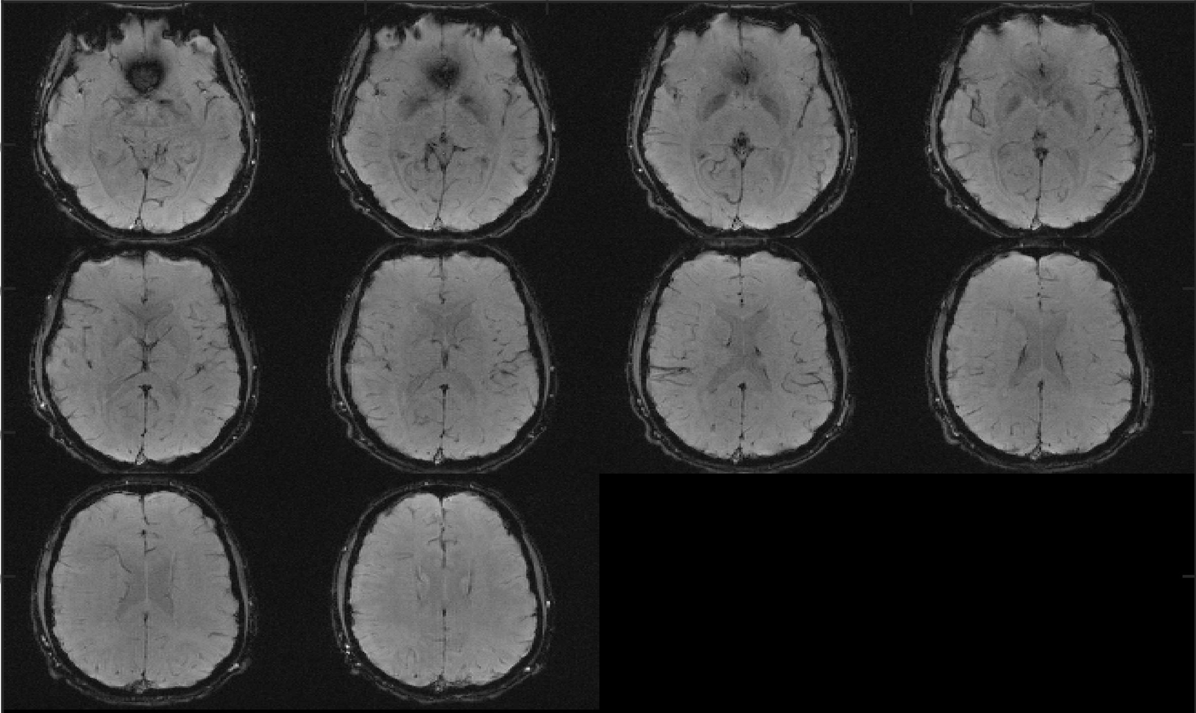
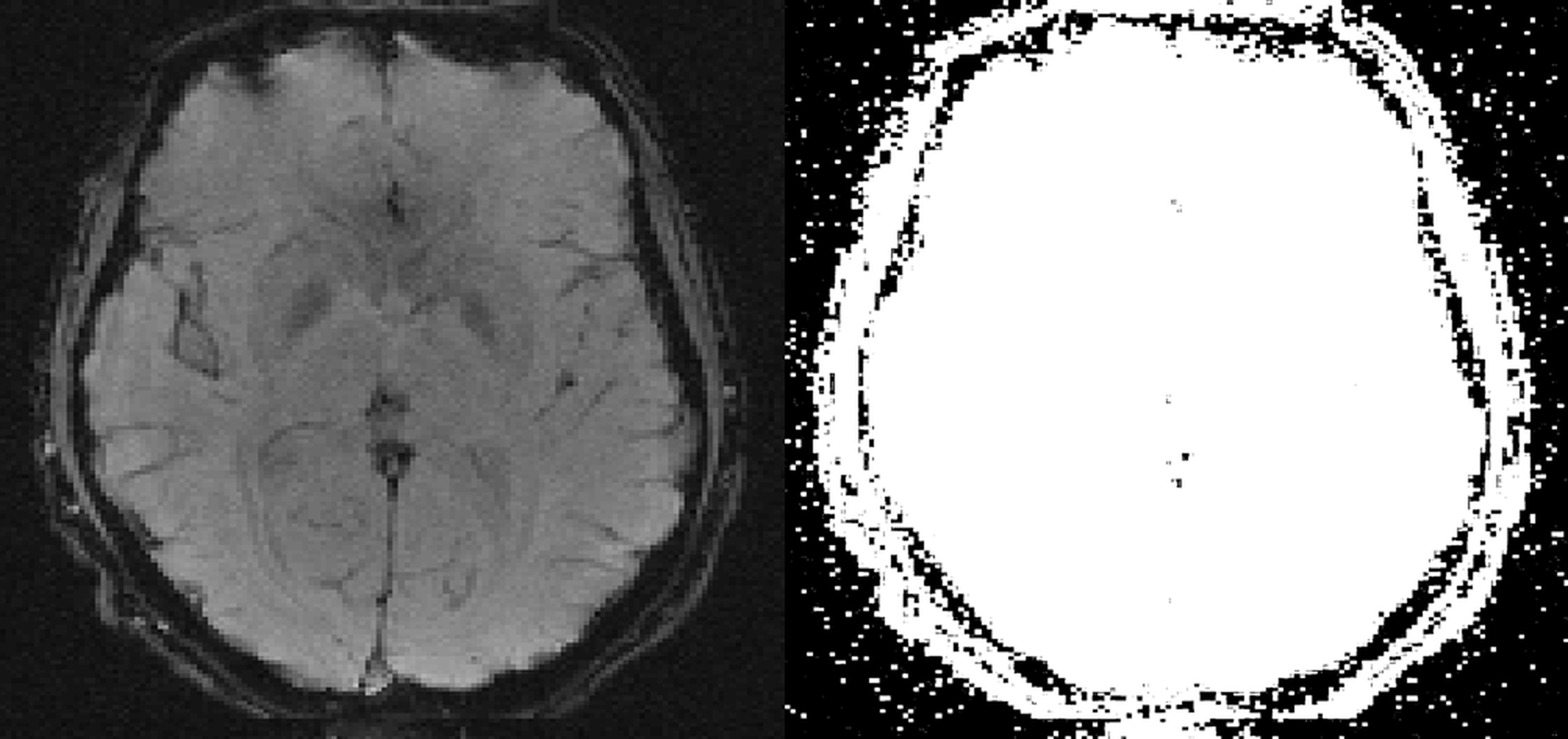
In Fig. 2 one complete volume and in Fig. 3 a single slice out of this volume is shown. Additional a scaled version with high contrast clearly demonstrates that there are no remaining folding artefacts visible. For this protocol the maximum number of slices is twelve. In comparison, the single-shot EPI sequence would allow for 33 slices within the same TR. However, the efficiency of VB-EPI, due to the nearly continuously sampling during RO is about 12% higher compared to standard blipped EPI.Discussion:
In the single slice image, anatomical structures, such as the optic radiation, the Putamen and the Globus Pallidus are clearly visible due to the high in-plane resolution of 1.2 mm. The limitation of this method is the relatively long TR, which results from acquiring the data with three RO-segments. So, this type of sequence will always require a factor of number of RO segments longer TR than a comparable single-shot EPI sequence with the same TE. This limitation can be addressed by Simultaneous Multi-Slice (SMS). Compared to single-shot EPI, the advantages of rs-EPI are reduced distortions and less blurring. This defines the scope of this sequence for high resolution, ultra-silent partial brain acquisitions. Increasing the duration of the PE blips to ½ echo-spacing, reduces acoustic noise at nearly no loss in image quality, even with standard Cartesian GRAPPA reconstruction. One would expect artefacts due to the non-Cartesian trajectory. However, as can be seen in Fig. 3 no folding artefacts are visible, which can be explained by the fact that the trajectory is only non-Cartesian at the edges of k-space. This can be addressed by skipping about 5% of the sampled data points at the edges of the readout. The resulting trajectory is very close to a Cartesian trajectory. However, the advantages of a lower acoustic noise and a higher efficiency are still preserved.Conclusion:
We have shown that it is feasible to acquire data with VB-EPI in combination with rs-EPI. This is highly interesting for quiet high resolution fMRI examinations. Furthermore, the data can be reconstructed with Cartesian parallel imaging methods without folding artefacts. The next step is to acquire fMRI data to validate the described benefits for fMRI.Acknowledgements
No acknowledgement found.References
1. Porter DA, Heidemann RM. High resolution diffusion-weighted imaging using readout-segmented echo-planar imaging, parallel imaging and a two-dimensional navigator-based reacquisition. Magn Reson Med. 2009;62(2):468-75.
2. Griswold MA, Jakob PM, Heidemann RM, Nittka M, Jellus V, Wang J, Kiefer B, Haase A. Generalized autocalibrating partially parallel acquisitions (GRAPPA). Magn Reson Med, 2002. 47(6): p. 1202-10.
3. Mansfield P. Multi-planar image formation using NMR spin echoes. J. Phys. C: Solid State Phys. 1977;10(3):L55
4. Cho ZH, Chung SC, Lim DW, Wong EK. Effects of the acoustic noise of the gradient systems on fMRI: a study on auditory, motor, and visual cortices. Magn Reson Med, 1998. 39(2): p. 331-5.
5. Schmitter S, Diesch E, Amann M, Kroll A, Moayer M, Schad LR. Silent echo-planar imaging for auditory FMRI. Magma, 2008;21(5):317-25.
Figures