5228
Functional MRI of the auditory cortex: maintaining the steady-state in EPI for silent inter-volume delays with different durations1Brain Imaging Center (BIC), Goethe University Frankfurt, Frankfurt am Main, Germany, 2Max Planck Institute for Empirical Aesthetics, Frankfurt am Main, Germany
Synopsis
An fMRI sequence for studying the auditory cortex is proposed, allowing for the insertion of silent delays of variable duration between EPI volumes during which auditory stimuli may be presented. Signal steady state conditions are maintained via spin saturation at fixed time points before each EPI volume. The method was successfully tested in vivo, yielding reliable activation of the primary auditory cortex. Results also show that high saturation efficiency is required to avoid erroneous activation patterns in compartments with long T1 such as cerebrospinal fluid.
Purpose
Investigations of the auditory cortex with functional MRI (fMRI) are hampered by acoustic noise during echo-planar imaging (EPI) acquisition [1]. To avoid acoustic interference in cognitive and systems neuroscience studies that investigate the perception of language and music, sparse sampling [2-3] is frequently employed, interleaving EPI acquisitions with silent periods for presenting sound stimuli. To ensure steady state conditions of the longitudinal magnetization, the inter-volume delays must be kept constant throughout the measurement which has been a serious constraint on sparse imaging for nearly the last two decades. The choice of a constant time interval between volumes is not necessarily an ideal solution for certain experiment designs. For example, sampling of the hemodynamic response curve may be facilitated by using variable delays during the measurement. Here, a novel acquisition scheme dubbed “pre-Saturated EPI using Multiple delays in Steady-state” (SEPIMS) is presented, allowing for the insertion of silent inter-volume delays with variable duration. Steady state conditions are maintained via robust spin saturation at fixed time points before each EPI acquisition. In this study, SEPIMS with two different inter-volume delays is tested and compared to conventional sparse sampling.Methods
Pulse sequence:
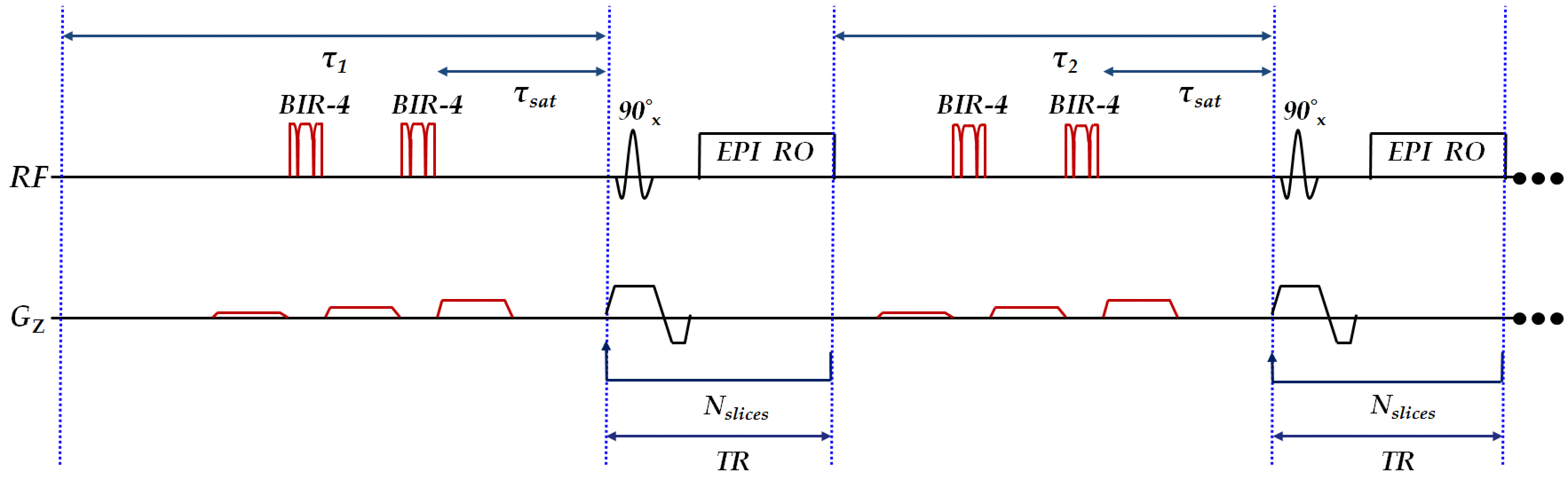
The SEPIMS sequence employed in this study (see Fig. 1) comprises two alternating silent periods τ1 and τ2 between successive EPI volumes. Spin saturation at a constant delay τsat before EPI acquisition yields identical longitudinal magnetization when acquisition starts and thus stable EPI signal levels. The pre-saturation module consists of two non-selective adiabatic saturation pulses BIR-4 [4], separated and surrounded by spoiler gradients which are relatively weak (1/2/4 mT/m), so concomitant acoustic noise is negligible. To avoid the risk of stimulated echo formation in the presence of long T2, e.g. in cerebrospinal fluid (CSF), spoiling across 8 subsequent silent periods is different by varying the gradient polarities. Pre-saturation comprises two BIR-4 pulses as demands on saturation efficiency are extremely high: signal variations due to incomplete saturation must be kept below the effect of interest.
Experiments:
The goal of this human fMRI experiment was to determine if SEPIMS is free from artifacts (in particular erroneous activation induced by residual signal variations), and yields similar results as a conventional sparse imaging technique. SEPIMS was tested on a healthy volunteer, using a 3T MRI-scanner with an 8-channel head receive coil and the parameters TR/TE: 2000/30ms, Field-of-view: 192×192mm2, spatial resolution: 2×2mm2, 30 axial-slices (thickness: 2mm, gap: 1mm), partial-Fourier: 6/8, GRAPPA-acceleration-factor: 2, echo-spacing: 0.87ms, Bandwidth: 1302Hz/pixel, τ1/τ2: 10/4s τsat: 3s, 48 volumes, duration 7:14min. Three experiments were performed, using (1) SEPIMS with double-saturation pulses, (2) SEPIMS with single-saturation pulses, (3) conventional sparse sampling with constant τ=12s and 32 volumes, duration 7:18min. Each experiment was repeated twice for improving the signal-to-noise ratio. An anatomical T1-weighted reference with an isotropic resolution of 1mm was also acquired.
Stimulus design for fMRI:
For activation of the auditory cortex in the superior temporal gyrus (STG), complex tone-bursts (30 pure tones ranging from 0.2 kHz to 6.4 kHz, amplitude-modulated in 5 Hz) with a duration of 6s were presented via MR-compatible headphones. For all three experiments, stimuli were presented before even volumes, only. Experiment 3 (conventional sparse sampling) was used as a reference study that can be expected to yield reliable activation in the STG.
Results and Discussion
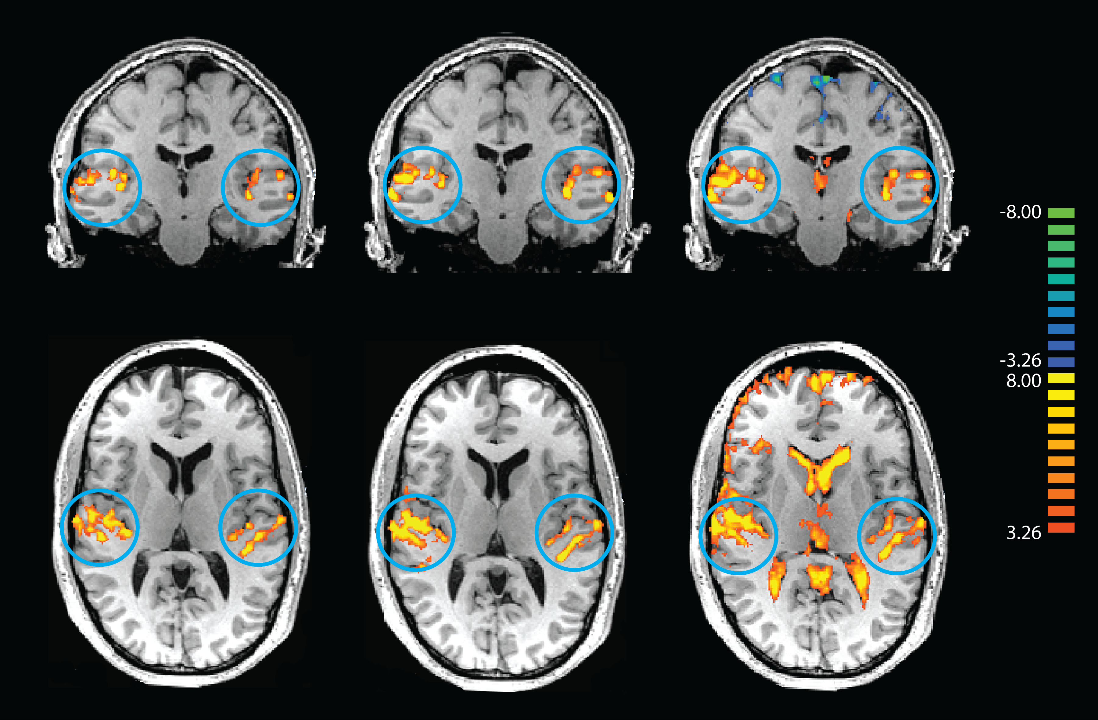
The conventional sparse sampling experiment yielded neuronal activation of the primary auditory cortex (Fig. 2, left). Comparable results were obtained with SEPIMS (Fig. 2, middle). However, SEPIMS with single saturation pulses yielded erroneous results in CSF of the ventricles and the sulci of the frontal cortex (Fig. 2, right). The results show that SEPIMS is well suited for auditory experiments with variable inter-volume delays. However, very high saturation efficiency is required since otherwise residual signal fluctuations may be misinterpreted as activation, in particular for compartments with long T1 values such as CSF.Conclusion
A novel fMRI acquisition scheme is proposed, allowing for silent inter-volume delays of variable duration. This permits flexible and efficient stimuli design in auditory fMRI experiments. The method was successfully tested in vivo, yielding reliable activation of the primary auditory cortex.Acknowledgements
No acknowledgement found.References
[1] Mansfield, Peter. "Multi-planar image formation using NMR spin echoes." Journal of Physics C: Solid State Physics 10.3 (1977): L55.
[2] Hall, Deborah A., et al. "Sparse temporal sampling in auditory fMRI." Human brain mapping 7.3 (1999): 213-223.
[3] Talavage, Thomas M., et al. "Quantitative assessment of auditory cortex responses induced by imager acoustic noise." Human brain mapping 7.2 (1999): 79-88.
[4] Staewen, R. Scott, et al. "3-D FLASH imaging using a single surface coil and a new adiabatic pulse, BIR-4." Investigative radiology 25.5 (1990): 559-567.
Figures