5217
High Resolution Pseudo Continuous Arterial Spin Labeling (pCASL) of mouse brain at 9.4 TeslaSankar Seramani1, Lydiane Hirschler2, Emmaneul Luc Barbier2, and Kuan Jin Lee1
1Laboratory of Molecular Imaging, Singapore Bioimaging Consortium, Singapore, Singapore, 2Université Grenoble Alpes, Grenoble Institut des Neurosciences, Grenoble, France
Synopsis
The objective of this study was to show the feasibility of performing ASL based high resolution perfusion imaging in the mouse brain at 9.4 Tesla. In this work we applied pCASL with phase optimization technique at the labeling plane to minimize the effect of B0 inhomogenity . We used fcFLASH based technique to measure the labeling efficiency of pCASL sequence and compared the labeling efficiency with FAIR ASL in mouse brain. Based on our results, with the proposed method of phase optimization, labelling slice can be place away from the iso-center of the magnet. This will allow us to place the imaging slice at the isocenter, which has showed significant improvement in the image quality of the mouse brain at ultra-high field strengths.
Introduction
The objective of this study was to show the feasibility of performing ASL based high resolution perfusion imaging in the mouse brain at 9.4 Tesla with improved image quality. pCASL is a modified form of CASL (Continuous Arterial Spin Labeling) where a short train of flow driven adiabatic pulses invert the spins in the feeding artery across the labeling plane. pCASL has been proven to show high SNR and sensitivity when compared to Pulsed ASL (PASL) techniques like FAIR. One of the main challenges in applying pCASL at Ultra High Field (UHF) is that the labelling efficiency is highly sensitive to the B0 Homogeneity at the labeling plane. In this work we applied previously reported pCASL with phase optimization technique [1] to measure perfusion on a mouse brain at 9.4 Tesla using phase optimization to minimize the effect of B0 inhomogeneity at the labelling plane. We used fcFLASH based technique to measure the labeling efficiency of pCASL sequence and compared the labeling efficiency with FAIR ASL in mouse brain.Materials and Methods:
MR Imaging experiments carried out in this study were approved and in compliance with IACUC (Institutional Animal Care and Use Committee). MR imaging was performed on 9.4T Bruker Biospec (Horizontal Bore) scanner/ Avance III Console/Paravision 6.01 interfaced with 86 mm volume transmit and 4-channel phased array receive coil. Two male Balb/C mice of twelve weeks old were used in this study. Animals were anesthetized with 3% isoflurane for induction and maintained at 2-3% isoflurane during the MR experiments. Respiration rate and body temperature were maintained at ~100 bpm and at ~37°C. One mouse was imaged twice, (one day interval).MR Imaging Experiments
The pCASL sequence and other labeling parameters are adapted from the previous reported article [1] on rat brain using unbalanced pCASL. The imaging slice was located at the iso-center and the labeling pulses were place at an angle perpendicular to the carotid artery approximately around 1cm away from the iso-center. 3D Mapshim procedure were done covering both the imaging and labelling slice before the imaging experiment. The labeling pulse train consisted of Hanning window shaped RF pulses with an B1 of 5μT, pulse duration/pulse rate= 400μs/800μs, Gmax/Gave= 45/5 mT/m, Both label and control phase optimization pre-scans were performed with the similar parameters with labeling duration τ= 1.5s, No of Repetition=1. The optimal labeling phase is computed and it was used to measure the actual pCASL perfusion measurement as reported earlier [1]. For the pCASL measurement, Labelling duration of τ=3s and Post Labelling Delay (PLD) of 300ms were used. Image acquisition was performed through single-shot EPI with FOV=2x2cm², slice thickness=1.5mm, matrix=96x96, TE=16ms, TR=4s, No of Repetition =30. To quantify CBF, T1 maps were acquired with single shot EPI with slice selective inversion at 10 TI’s ranging from 30 to 8000 with a TR of 10000) and inversion efficiencies were measured with fcFLASH sequence at ~4 mm above the labeling slice with PLD=0 and τ =200ms. A FAIR based PASL dataset was also measured with 30 TI’s and a shim volume covering from head to abdomen. The FAIR-derived CBF and labeling efficiency were compared with that of the fcFLASH based method. Perfusion imaging experiments were done on one mouse on different days to determine the repeatability of CBF derived from pCASL.Results and Discussion:
Figure
1(a)-1(f) shows the perfusion imaging results from a mouse performed on
different days, with same experimental setup and procedures performed during
the two sessions of the experiments. Table 1 shows the CBF results of the two
experiments on different regions (Cortex, Hippocampus & Thalamus) of the
mouse brain. Our results of mouse brain CBF are in agreement with the
previously published article [2] at 11.7 Tesla by Duhamel et al. The reproducibility of the two experiments conducted on the
same mice shows high reproducibility of pCASL sequence in measuring the CBF.
Conclusion:
Based on our results, with the proposed method of phase optimization, labelling slice can be place away from the iso-center of the magnet. This will allow us to place the imaging slice at the iso-center, which has showed significant improvement in the image quality when compared to Duhamel et al [2] of the mouse brain at ultra-high field strengths.Acknowledgements
No acknowledgement found.References
Reference: [1] Hirschler et al, Proc of ISMRM 2015;[2]Duhamel et al, Magn Reson Med 2014;Figures
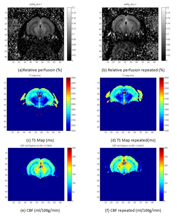
Figure
1: pCASL based Cerebral Blood Flow(CBF, in ml/100g/min) measurement for control
mouse
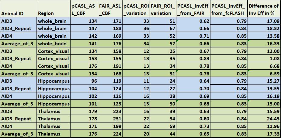
Table 1: Results of CBF in the mouse brain using PCASL and
FAIR.