5215
High resolution PET insert for high field preclinical MRI: evaluation of single ring system using 7T field strength1Biomedical MRI, Department of Imaging and Pathology, KU Leuven, Leuven, Belgium, 2Nuclear Medicine, Department of Imaging and Pathology, KU Leuven, Leuven, Belgium, 3i3M, Valencia, Spain, 4Oncovision, Valencia, Spain, 5Bruker Biospin, Ettlingen, Germany
Synopsis
We designed a novel PET insert based on monolithic LYSO crystals. From our first evaluation, we can conclude that sub-millimeter detector spatial resolution, combined with accurate photon DOI determination, make it possible to acquire high resolution reconstructed images. This enables us now to combine simultaneously high resolution and sensitivity PET with high field preclinical MRI to extract simultaneously complex data from anatomical to molecular information and to dynamically follow non-invasively animal models of different pathologies with no compromise in performance of each imaging modality.
Introduction
Methods
The
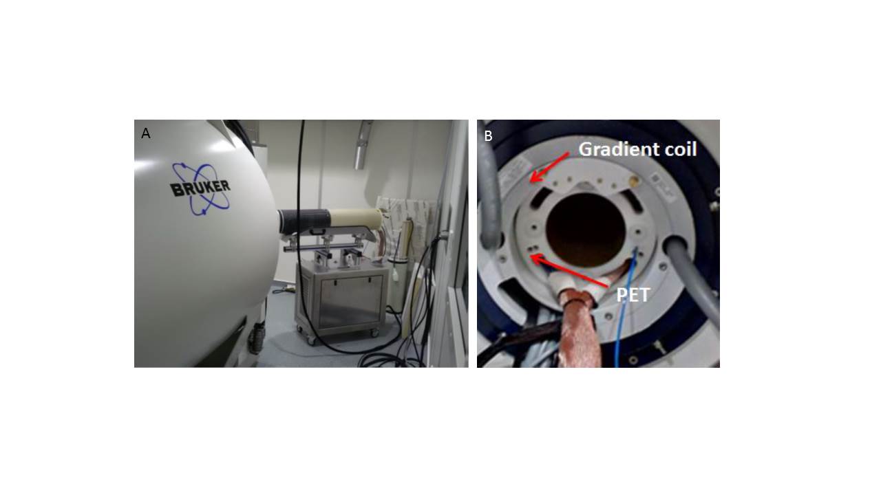
PET system formed by 8 detectors has been tested inside the Bruker BioSpec 70/30
USR with a magnetic field of 7 T (Figure 1). The MR system was equipped with a
BGA 20S-HP gradient coil. The inner and outer diameters of the PET scanner
(insert) are 114mm and 198mm, respectively, fitting inside the MR gradient. Quadrature
birdcage coils were used for all experiments (86mm inner diameter for phantom
and rat studies and 40mm for mouse imaging). For the RF shielding, we
implemented Carbon Fiber structures with tubular shape, surrounding the PET
electronics. Fast Spin echo and EPI sequences were tested simultaneously with
the PET insert. We assessed potential eddy currents induced by fast switching
of the gradient, field homogeneity through B0 maps and PET/MR image
quality (resolution, SNR, image homogeneity etc.). For PET characterization, we
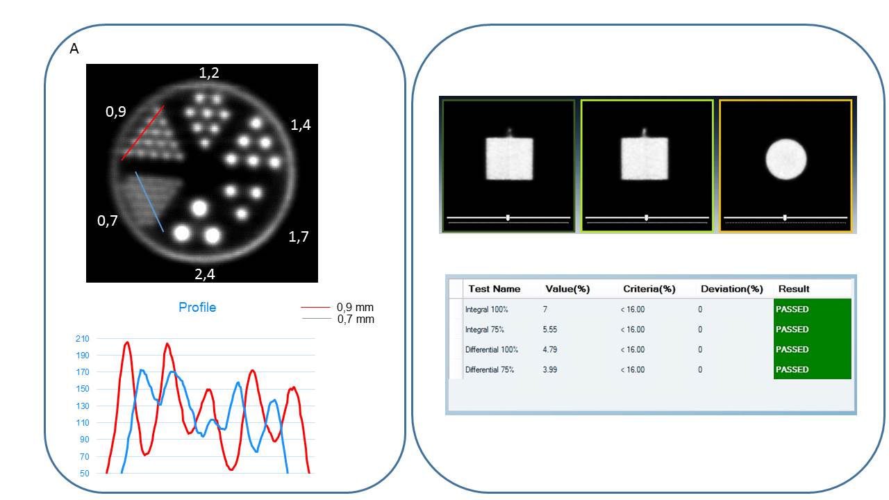
followed the NEMA protocol for sensitivity, and use mini-Derenzo like phantom
(filled with 150uCi of 18F-FDG) for estimation of resolution and
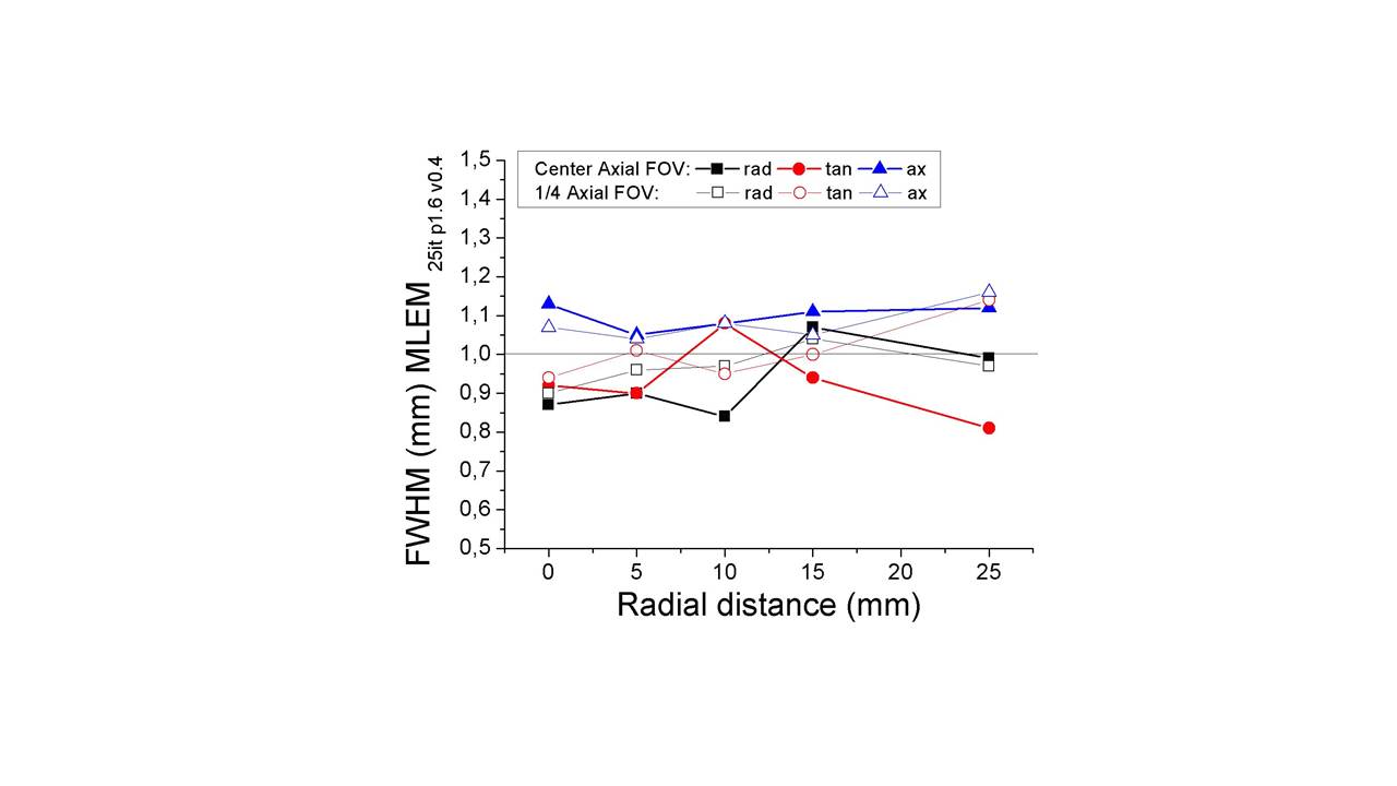
image quality. For all tests, PET data were reconstructed using Maximum
Likelihood Estimation Method (MLEM) with either voxel size of 0.5 or 0.29mm3
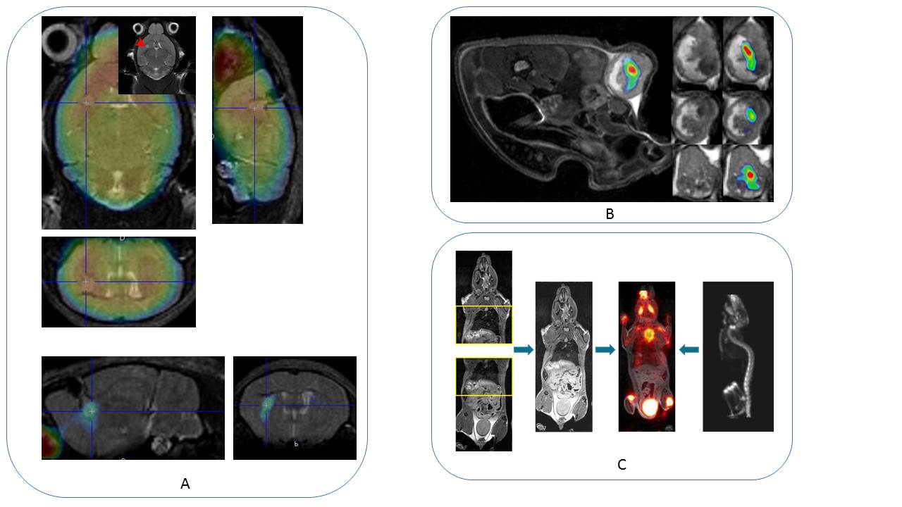
and at least 12 iterations. Several animal models (mouse glioma, mouse
stroke, xenografts in mice, rat heart, see Figures) were evaluated to provide
real in-vivo data for the quality assessment of the simultaneous PET/MRI
acquisitions.Results
We
tested different RF pulses (20ms and 630W, with 51us and 1ms duration) and MRI
sequences (RARE, EPI, etc…) without observing PET degradation of the PET image
quality or eddy currents that could produce a sub-optimal MR performance. The
FieldMap sequences showed in the present study no change in the B0 field with a
55mm spherical phantom when the PET insert was inside the MR scanner (SNR
variation with/without PET <6%). Both RARE and EPI sequences showed ghost
levels of about 2.4 to 3.6%. The PET geometry and performance were almost
identical to the current in-line Albira Si system6. Sub-millimeter image
resolution (between 0.9 and 0.7mm) and homogeneous-FOV spatial resolution were
reached, as shown in Fig.2 and Fig.3. The sensitivity for the one-ring PET,
following the NEMA protocol, was determined to be beyond 3.5%. In-vivo evaluation demonstrated the addedd value
of using simultaneously high resolution PET and MRI (Fig. 4). For example, a glioma
as small as 0.6mm3 were visualized on the MRI and small SUV
differences with contralateral side were demonstrated by PET. Dynamic data were
acquired using cardiac PET and MRI acquisition in the rat heart.Discussion and Conclusion
The design of a first prototype of a
small animal PET insert is finalized and was successfully tested within a 7T
MRI (Bruker Biospec). Typical MR image sequences for anatomical and functional
imaging did not affect the PET performance. In-vivo experiments demonstrated
the benefit of such combination in small animal models.
PET detector spatial
resolution nearing 1mm, combined with accurate photon DOI determination, make
it possible to return high resolution reconstructed images visualizing the 750um
rods of the micro Derenzo-like phantom. This enables us to combine high
resolution and sensitivity PET with high-field preclinical MRI to extract
simultaneously complex data from anatomical to molecular information and to
dynamically follow non-invasively animal models of different pathologies.
Acknowledgements
MRI and PET imaging systems were founded by the Hercules foundation (PI: U. Himmelreich) and Stichting tegen Kanker (PI: C. Deroose)References
1Schug D, Lerche C, Weissler B, et al. Initial PET performance evaluation of a preclinical insert for PET/MRI with digital SiPM technology. Phys Med Biol. 2016 Apr 7;61(7):2851-78.
2Von Schulthess G K and Schlemmer H P W. A look ahead: PET/MR versus PET/CT. Eur. J. Nucl. Med. Mol. Imaging. 2009, 36 3-9.
3Drzezga A et al. First clinical experience with integrated whole-body PET/MR: comparison to PET/CT in patients with oncologic diagnoses J. Nucl. Med. 2012, 53 845–55.
4Jadvar H. and Colletti P. M. Competitive advantage of PET/MRI Eur. J. Radiol 2014. 83 84–94.
5Vandenberghe S. and Marsden P. K. PET-MRI: a review of challenges and solutions in the development of integrated multimodality imaging. Phys. Med. Biol. 2015, 60 R115.
6Gonzalez A.J., et al., Trans. Nucl. Science 63, pp. 2471, 2016.
Figures