5197
Flexible spin echo triple echo Dixon (flexible STED) for fat suppressed T1-weighted imagingJong Bum Son1, Colleen Costelloe1, Ken-Pin Hwang1, Tao Zhang2, Ersin Bayram2, John Hazle1, and Jingfei Ma1
1The University of Texas MD Anderson Cancer Center, Houston, TX, United States, 2GE Healthcare, WI, United States
Synopsis
The conventional spin-echo (SE) sequence produces the best available T1-contrast and is often used for imaging of brain, head and neck, and spine. Unfortunately, the inclusion of fat suppression pulses in an SE acquisition substantially reduces the number of slices per TR and therefore the overall scan efficiency. In this work, we developed a flexible spin-echo triple-echo Dixon (flexible STED) technique that enables SE Dixon imaging with high efficiency. The feasibility of the technique is demonstrated with in vivo post-contrast fat-suppressed T1-weighted imaging of spine.
INTRODUCTION
Compared to most other pulse sequences for T1-weighted imaging (e.g., spoiled gradient echo, FSE,T1FLAIR), the conventional spin-echo (SE) sequence produces the best available T1-contrast and is often used for imaging of brain, head and neck, and spine. Unfortunately, the inclusion of frequency-selective fat suppression (CHESS-type) pulses in an SE acquisition substantially reduces the number of slices per TR and therefore the overall scan efficiency. Previously, an interleaved two-point Dixon SE sequence has been shown to produce much improved fat-suppressed T1-weighted images at 3T over fat-suppressed T1-weighted images by SE with CHESS [1]. The purpose of this work was to develop a flexible spin-echo triple-echo Dixon (flexible STED) technique that is more efficient and suitable for fat-suppressed T1-weighted imaging. The feasibility of the technique is demonstrated with in vivo post-contrast T1-weighted imaging of spine.METHODS
Fig. 1 shows the proposed flexible STED pulse sequence. The technique is an SE implementation of the previously published flexible fast spin-echo triple-echo Dixon (FTED) sequence [2]. In flexible STED, the single readout gradient in conventional SE is replaced with three fast-switching bipolar gradients (gxw-, gxw, and gxw+) to produce three echoes. The middle echo (gxw) is placed at the conventional spin-echo position. The first (gxw-) and the third (gxw-) echoes are separated from gxw by an inter-echo-spacing (). The pulse sequence requires no deadtime between the echo readouts except for the gradient switching. Therefore is determined by the readout durations and can be flexible depending on the receiver bandwidth (RBW) and the matrix size along the readout direction. The three echoes are identically phase encoded to produce three raw images (S-, S, and S+) with the relative water and fat phase shifts of -θ, 0, and +θ, respectively. As in flexible FTED, these three raw images are fed into a jointly processed two-point Dixon algorithm to produce a combined water-only and fat-only image for each slice [3]. We implemented the flexible STED sequence on a GE 3T whole-body MRI scanner on the HDxt 16.0 platform (GE Healthcare, Waukesha). Written informed consent was obtained for the study and sagittal spine images using the flexible STED were acquired using a 6-channel spine coil (GE Healthcare, Waukesha) after injection of a contrast agent (Gadovist, Bayer, Berlin). The scan parameters were: TR/TE = 700/11ms, acquisition matrix = 320 x 448, FOV = 28 cm x 28cm, RBW = ±200 kHz, slice thickness/gap= 3/1 mm, and a total of 15 slices were acquired in 5:24 mins. The inter-echo spacing was 1440 ms for these scan parameters, corresponding to a water and fat relative phase angle of 154°. The maximum number of 25 slices were allowed without an increase to the scan time. In comparison, a maximum number of only 20 slices would be allowed if SE with ChemSat were used with otherwise identical scan parameters. As part of the clinical protocol, we also acquired post-contrast T1-weighted images of the same patient with T1 fluid-attenuated inversion recovery (T1FLAIR) with fat suppression. The scan parameters were identical except for ETL = 8, TR/TE = 3000 ms/24 ms, RBW = ±50 kHz, and scan-time = 4 mins 20 secs. The algorithm used for processing the flexible STED images was implemented in MATLAB (MathWorks, Natick, Massachusetts, USA).RESULTS
Fig. 2a shows a sagittal fat-suppressed post-contrast T1-weighted image by T1FLAIR. In T1FLAIR, fat suppression was achieved with a spectrally selective inversion recovery RF pulse and fast spin-echo acquisition (with an ETL of 8) was used to make the scan time clinically feasible. Fig. 2b) shows a corresponding water-only image by the flexible STED sequence, which is spin-echo based and uses Dixon processing to achieve fat suppression. The triple-echo readout in the flexible STED enabled the acquisition of fat-suppressed T1-weighted SE images with high efficiency. Noticeable differences can be appreciated in the image contrast and contrast enhancement between the two different T1-weighted images.DISCUSSION
The flexible FTED sequence [2] has been shown to be useful for T2-weighted imaging with uniform fat suppression. The proposed flexible STED technique is an extension of the flexible FTED and makes it clinically feasible to perform fat suppressed T1-weighted imaging with a short TR SE acquisition. Since STED avoids the use of refocusing echo trains, T1 weighting is improved over FSE-based methods.Acknowledgements
Part of the research was conducted at the Center for Advanced Biomedical Imaging at The University of Texas MD Anderson Cancer Center with equipment support from GE Healthcare.References
[1] Ma J, Hwang K-P, Ginsberg L. Improved image contrast and scan efficiency for fat suppressed T1weighted imaging at 3T with a spin echo two-point Dixon technique. Proc Intl Soc Magn Reson Med, 5/2010. [2] Son, JB, Hwang KP, Madewell JE, Bayram E, Hazle JD, and Jingfei Ma. A Flexible Fast Spin Echo Triple-Echo Dixon Technique. Magn Reson in Med. 2016; DOI 10.1002/mrm.26186. [3] Ma J, Son JB, Hazle JD. An improved region growing algorithm for phase correction in MRI. Magn Reson in Med. 2015; DOI 10.1002/mrm.25892.Figures
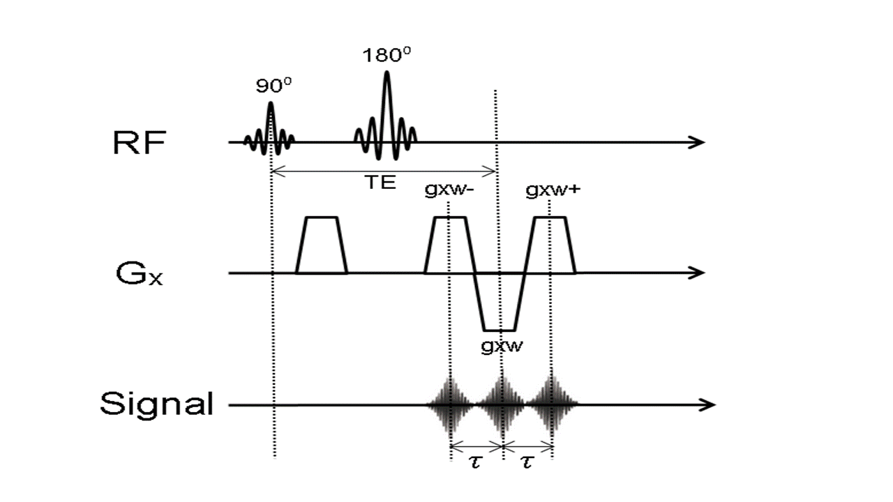
Fig. 1. Pulse
sequence diagram of the proposed flexible STED technique.
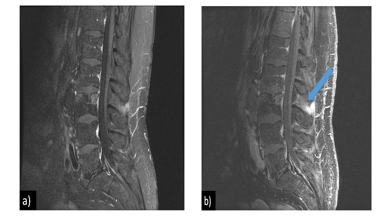
Fig. 2. In vivo
post-contrast fat-suppressed sagittal T1-weighted spine images (a) by T1 FLAIR
(b) by the proposed flexible STED. Block arrow indicates an area of different
contrast enhancement.