5187
In-Vivo Fat Water Separation with Multiple-Acquisition bSSFPMichael Anthony Mendoza1, Joseph Valentine2, and Neal Bangerter3
1Electrical Engineering, Brigham Young University, North Ogden, UT, United States, 2Electrical Engineering, Brigham Young University, provo, UT, United States, 3Electrical Engineering, Brigham Young University, Provo, UT, United States
Synopsis
In this work, we present a novel technique that combines the advantages of bSSFP with Dixon reconstruction in order to produce robust water fat decomposition with high SNR in a short imaging time, while simultaneously reducing banding artifacts that traditionally degrade image quality.
Introduction
Balanced Steady State Free Precession (bSSFP) is a fast acquisition technique with high signal-to-noise ratio (SNR), but is sensitive to off-resonance effects that cause signal modulation and banding artifacts. These bands of signal loss degrade image quality. Typically, band reduction is achieved by combining multiple RF phase cycled bSSFP images that have spatially shifted bands. Dixon reconstruction methods use a linear combination of ‘in-phase’ and ‘out-of-phase’ images to extract separate water and fat images. This work is a continuation of previous efforts to combine Dixon reconstruction with multiple acquisition bSSSP to give a more robust water fat separation with high SNR in a short imaging time, while simultaneously reducing the banding artifacts typically found in SSFP. 1 Specifically, we have improved the algorithm by incorporating phase unwrapping, a field map, and other improvements and demonstrate results in-vivo.Theory
At large flip angles, bSSFP images have a sinusoidal-like shaped spectral profile with a pass band region and a region of signal null. These profiles are a function of off-resonance frequency and can be shifted using RF phase cycling. Four phase cycled bSSFP images with delta phi = 0°, 90°, 180°, 270° generates an evenly distributed set of signals along a bSSFP spectrum. Using a nonlinear averaging reconstruction, these images are combined to reduce banding artifacts.3, 4 The choice of echo time (TE) determines the relative phase between water and fat components. These four phase cycled images are acquired at different echo times to generate ‘in-phase’ images and ‘out-of-phase images. Clever linear combinations of these images that avoid signal nulls are used to produce separate water and fat images. A phase map of the magnetic field inhomogeneities is generated from the acquired images, and used to improve the reconstruction.Methods
Four phase-cycled bSSFP axial images of a knee and a pelvis were acquired using a 3T Siemens scanner. These images were collectively generated with a flip angle of 90 degrees and a TR of 10ms. Each image was phase cycled with delta phi = 0°, 90°, 180°, 270° and TEs = 5ms, 6.17ms, 5ms, and 6.17ms respectively. Images were reconstructed on each voxel independently.Results & Discussion
The results for the axial knee acquisition are shown in Figure 1, and those for the pelvis are shown in Figure 2. On the left in each set of images are a single phase-cycled bSSFP image (exhibiting banding artifact) as a reference. The middle images of each are fat only, and the right images are water only. Note the ability of the algorithm to simultaneously separate fat and water while reducing banding artifacts. These results are in good agreement with theory and show the algorithm is capable of in-vivo water fat separation. Future work will focus on improving the performance of the algorithm in cases of partial-voluming of fat and water, the use of region-growing algorithms to better handle water and fat boundary transitions, and better incorporation of phase map data.Acknowledgements
Thanks for all your help Joe Valentine and Dr. Bangerter.References
[1] Mendoza et al., ISMRM 22nd Annual Meeting 2014
[2] Quist et al., Magnetic Resonance in Medicine, 2012
[3] Elliot et al., Magnetic Resonance Imaging, 2007
[4] Bangerter et al., Journal of Magnetic Resonance in Medicine, 2004;
[5] Glover and Schneider, Journal of Magnetic Resonance in Medicine, 1991
[6] Dixon, Journal of Radiology, 1984
Figures
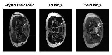
Example bSSPF phase cycled image (left), Water reconstructed images (center), Fat reconstructed image (right)
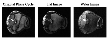
Example bSSPF phase cycled image (left), Water reconstructed images (center), Fat reconstructed image (right)