5173
Spiral SENSE MP-RAGE using long readouts and an expanded signal model1Institute for Biomedical Engineering, ETH and University of Zurich, Zurich, Switzerland
Synopsis
Spiral readout was used to accelerate the standard T1-weighted 3D head scan based on MPRAGE at 3 Tesla without any compromise on images quality. The encoding model used by the reconstruction included the static magnetic field map as well as the k-space trajectory and a dynamic field correction provided by a field camera. The study demonstrates the utility of state-of-the-art spiral scanning for routine MRI applications.
Introduction
Spiral trajectories have long been used in ultra-fast MRI for applications requiring short scan time, short TE and reduced motion artefacts. However, the propagation of this efficient technique to the routine radiological practice has not started until recently (1). This is due to several technical challenges such as the increased sensitivity to static- and time-dependent magnetic field perturbations and the complexity of combining gradient- and sensitivity encoding. Currently, all these problems can be handled efficiently by field mapping/monitoring and proper reconstruction techniques. We demonstrate that the standard protocol for a 3D T1-weighted head scan based on MP-RAGE (inversion recovery) can be accelerated using spiral trajectories without compromise on image quality.Methods
The sequence was based on an interleaved 2D gradient echo spiral with phase encoding of the slab dimension (cylindrical stack of spirals) combined with non-selective inversion-recovery preparation. Following an adiabatic inversion RF pulse, several low-flip angle slab-selective excitations of the same spiral segment and different values of slab-encoding were applied. This sequence was repeated in two nested loops, the inner one running over slab-encoding segments, and the outer over spiral interleaves. This loop structure, which could be implemented by parametrizing manufacturer’s standard software, assures that all spiral interleaves in one stack plane have the same recovery time (TI) and thus identical starting signals. The variation of TI is mapped solely to the linearly sampled slab dimension as in the standard Cartesian MPRAGE, which leads to a smooth k-space weighting and avoids ghosting artefacts. The effective TI is defined by the segment that hits the k-space origin in the slab dimension. The experiment was carried out on a 3 Tesla MRI system (Achieva, Philips, The Netherlands) equipped with an 8-channel head array and 30 mT/m gradients. The magnetic field was monitored concurrently using a 16-probe field camera (Scope, Switzerland), which provides information about fluctuations of B0, k-space trajectory and field components up to 3rd order (2). A double echo sequence was used to measure the static field map and receiver array sensitivities. Both the static map and the monitored dynamic field were used for the image reconstruction based the iterative method using the expanded signal model (3). The proof-of-principle study presented here used 1x1 mm resolution in-plane and 2mm in slab direction, and contained 16 spirals per inversion recovery. With 20 spiral interleaves, 20ms acquisition window length, TR=4s, and TI=1.3S the total scan time was 2min 40 sec. The scan was repeated with two echo time values to allow fat/water separation using the Dixon 2-point method.Results and discussion.
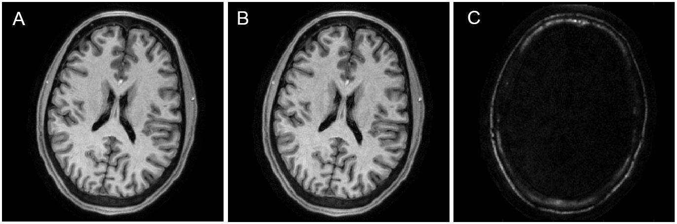
Figure 1 shows water and fat images of the central slab plane obtained from the full-sampled data. A water image reconstructed from one-half of spiral interleaves using non-cartesian SENSE (4) is shown Fig. 1. C. No artefacts due to resonance offset or time-dependent field perturbations are visible. There are no errors of water/fat separation. This is possible with only two echo times because the reconstruction, which includes the field map in the expanded signal model, effectively removes the off-resonance phase deviations. This initial study used an acquisition duty cycle of less than 50% of what the gradient system allows (50ms repetition rate of 20ms-long spiral scans). Still, a scan time reduction factor of about two could be reached compared to a standard Cartesian scan. By increasing the excitation rate and taking more spirals per inversion, a two- to 4-times higher number of encoding steps in the slab direction should be acquired without time penalty. We aim at a total scan time below 2 minutes for a 1-mm isotropic resolution MPRAGE scan of the whole brain and a comparable speed gain with other standard MRI sequences.Conclusion
Spiral k-space trajectories with readout times as long as 20ms can be used for high resolution MRI at 3T with clinically useful image quality. The sampling efficiency of long-readout spirals allows a significant reduction of the scan time.
Acknowledgements
No acknowledgement found.References
1. Pipe JG et al, Progress towards Robust Spiral MRI for Rapid Brain Exams, Proc. ISMRM 2016, p 88
2. Wilm BJ et al. Higher order reconstruction for MRI in the presence of spatiotemporal field perturbations. Magn Reson Med 65 (2011): 1690-1701.
3. Barmet C, De Zanche N, and Pruessmann KP, Spatiotemporal magnetic field monitoring for MR, Magn Reson Med 60 (2008): 187-197.
4. Pruessmann KP, Weiger M, Börnert P and Boesiger P, Advances in sensitivity encoding with arbitrary k-space trajectories, Magn Reson Med 46 (2001): 638-651.
Figures