5172
Enhancing resolution in single-shot MRI by SUper-resolved SPEN with SENSE (SUSPENSE)1Chemical Physics, Weizmann Institute of Science, Rehovot, Israel, 2Department of Electrical Engineering and Computer Sciences, Berkeley, CA
Synopsis
Spatio-temporal encoding (SPEN) delivers single-scan images with increased robustness to shift and susceptibility artifacts. These acquisitions are usually carried out in a “hybrid” mode that prevents a sufficiently dense sampling along the SPEN domain. Alleviating this resolution loss had so far demanded the acquisition of multiple interleaved scans. The present study demonstrates that by relying on multiple sensors, a similar resolution enhancement can be achieved in a single shot. The principles and potential of the ensuing Super-resolved SPEN with SENSE (SUSPENSE) is demonstrated, with sub-mm single-shot 3T image acquisitions on phantoms and humans.
Motivation
Single-shot 2D SPEN is carried out in a hybrid mode, where “blips” sample the low-bandwidth domain in image space. In most instances a sufficiently dense sampling of the SPEN domain is unfeasible, but the ensuing loss in spatial resolution can be offset by the acquisition of multiple interleaved scans [1]. Unlike EPI interleaving, where the aim of the blips is to increase the sampling density so as to cover the FOVy without folding artifacts [2], SPEN interleaving samples regions in space that had been skipped by the original rasterization. Still, whether sampling ky or y, interleaved EPI and SPEN share the common need for performing multiple scans. Techniques such as SMASH/SENSE/GRAPPA can be used to fill missing lines in k-space [3-5] –either alone or in combination with compressed-sensing (CS) [6-7]. The present study exploits this to achieve a resolution enhancement that is identical to that afforded by interleaved SPEN, but can be completed in a single scan.Theory
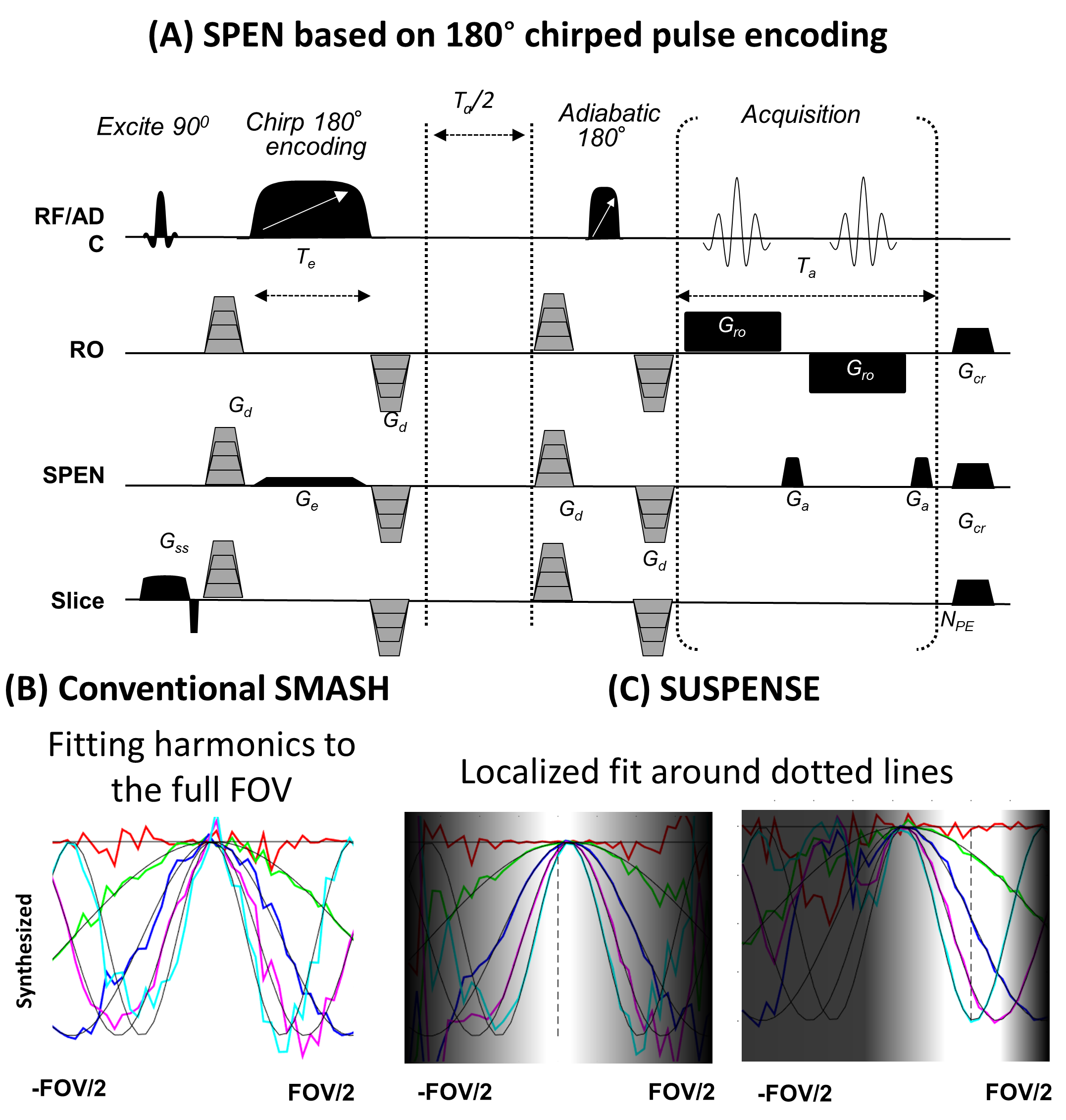
The SPEN pulse sequence used in this work (Figure 1) imparts a parabolic phase on the spins along their y axis. By spatially advancing this parabola, the acquisition gradient Ga manages to image the full FOVy. Ideally, Ga blips would be fine enough to maximize the ensuing image resolution, which for fully-refocused SPEN [8,9] demands blipping $$$N^{max}_{SPEN}=2\gamma G_aFOV_yT_e$$$ times [2,10]. While this is hard to achieve in a single shot, multiple sensors can make up for this deficit. To visualize this notice that although they are implemented for different purposes, both SPEN and EPI interleaving premodulate acquisitions by a certain factor $$$\exp[-iΔk(l-1)y]$$$, over $$$1 ≤ l ≤ Nshot$$$ interleaved scans. According to the SMASH formalism [3] such spatial phase variations can, if sufficiently slow, be mimicked by summing signals arising from multiple coils: $$$\exp[-ih\Delta k\cdot y] =\sum_cn^h_cS_c(y)$$$ , where $$$n^h_c$$$ are suitable weighting coefficients for harmonic h and $$$S_c$$$ are sensitivity maps for the various c-coils. Such formalism is usually of limited usefulness since the weighting coefficients need to hold throughout the entire FOVy (Figure 1B); in SPEN however, where signals stem from a localized spatial neighborhood, it is simple and robust to extend such formalism for the sake of computing the “missing” interleaved data. To do so we look for a RO and SPEN (x and y) set of localized coefficients , such that the coils will satisfy, for that particular y-neighborhood, $$\sum_{c=1}^{n_c}n^h_{c,y}W(y)S_c(x,y) = W(y)\exp[-ih\Delta k\cdot y],\ \ \ y\in FOV_y$$ where Nc is the number of coils and $$$W(y)$$$ is a weighting function which emphasizes the interpolated location. This enables one to faithfully synthesize localized harmonics up to a high resolution-enhancement factor R (Figure 1C). It is feasible to incorporate into this formalism, SENSE- and GRAPPA-based reconstruction algorithms. In the former case one can also search numerically for an image I satisfying A(I)=Data, where A is an operator which includes the quadratic phase and the sensors’ sensitivities. This was here implemented using CS reconstruction with a total-variation filter [7], leading to Super-resolved SPEN with SENSE (SUSPENSE). SUSPENSE, like SMASH, requires a coil sensitivity map. The direct-space sampling of SPEN, however, implies that despite its undersampling, unfolded low-resolution images carrying their respective sensitivity maps will arise; this renders the method completely self-referenced and fully auto-calibrated. Additionally, and also unlike the EPI case, the R-factor can be set in post-processing; the maximal R is limited by the sensors coverage over the FOV, and should be limited to $$$N^{max}_{SPEN}/Npe$$$.Methods
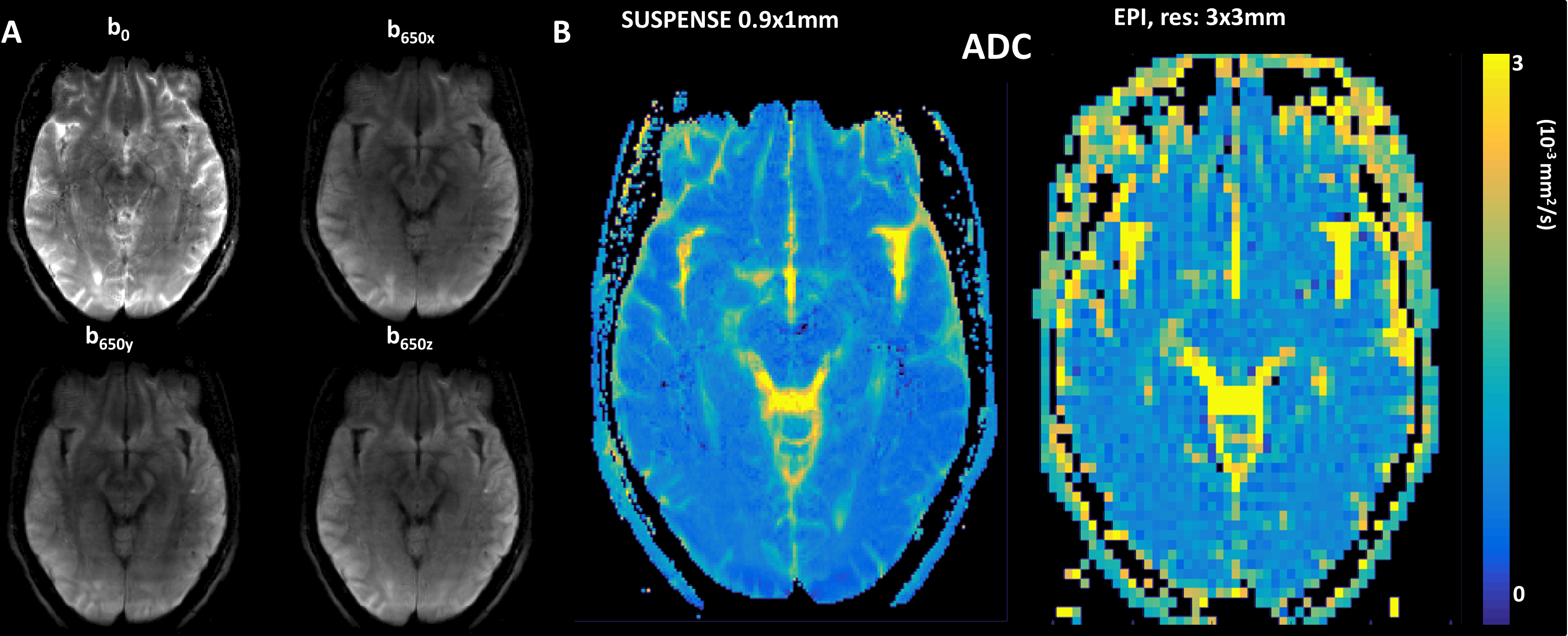
SUSPENSE was validated on a phantom with a stripe pattern of sub-mm separation and on healthy human volunteers, using a 3T Siemens with a 32-channel head coil. Applications involved DWI imaging at sub-mm resolution.Results and Discussion
Figure 2 shows phantom image reconstructions performed for increasing R-factors. In the original single-shot resolution (0.94mm) the phantom stripes are blurred, but higher Rs succeed to resolve these pattern. Notice that R-factors ≥3 do not increase the effective resolution, as with it $$$N^{max}_{SPEN}$$$ has been reached. Figure 3 shows a DWI experiment acquired at a native in-plane resolution of 4.5x1mm (SPENxRO) with a high $$$N^{max}_{SPEN}=200 $$$ value (FOV=18cm). Reconstruction using SUSPENSE with R=5 results in a final 0.9x1mm in-plane resolution. The high bandwidth used provides high robustness to inhomogeneities, otherwise noticeable at the frontal tissue-air border region.Conclusions
SUSPENSE provides robust, high resolution anatomical and ADC maps, free from multi-shot complications.Acknowledgements
Funding by grants ISF 795/13, ERC-2014-PoC # 633888, Minerva Foundation #712277, and the Kimmel Institute of Magnetic Resonance (Weizmann), is acknowledged. ML acknowledges a Visiting Faculty Program Fellowship (Weizmann). We are also grateful to Dr. Sagit Shushan (Wolfson Medical Center), and the Weizmann MRI team (Edna Furman-Haran, Fanny Attar and Nachum Stern).References
[1] Interleaved multishot imaging by spatiotemporal encoding: A fast, self-referenced method for high-definition diffusion and functional MRI. Schmidt R, Seginer A, Frydman L. Magn Reson Med. 2016 May;75(5):1935-48
[2] Interleaved echo planar imaging on a standard MRI system. Butts K, Riederer SJ, Ehman RL, Thompson RM, Jack CR. Magn Reson Med. 1994 Jan;31(1):67-72.
[3] Simultaneous acquisition of spatial harmonics (SMASH): fast imaging with radiofrequency coil arrays. Sodickson DK1, Manning WJ Magn Reson Med. 1997 Oct;38(4):591-603
[4] SENSE: sensitivity encoding for fast MRI. Pruessmann, K.P., Weiger, M., Scheidegger, M.B. and Boesiger, P., Manning WJ Magn Reson Med. 1999, 42(5), pp.952-962.
[5] Generalized autocalibrating partially parallel acquisitions (GRAPPA). Griswold MA, Jakob PM, Heidemann RM, Nittka M, Jellus V, Wang J, Kiefer B, Haase A Magn Reson Med. 2002 Jun;47(6):1202-10
[6] ESPIRiT—an eigenvalue approach to autocalibrating parallel MRI: where SENSE meets GRAPPA. Uecker M, Lai P, Murphy MJ, Virtue P, Elad M, Pauly JM, Vasanawala SS, Lustig M Magn Reson Med. 2014 Mar;71(3):990-1001.
[7] Sparse MRI: The Application of Compressed Sensing for Rapid MR Imaging. M. Lustig, D.L Donoho and J.M Pauly, Magn Reson Med. 2007 Dec; 58(6):1182-1195
[8] RASER: a new ultrafast magnetic resonance imaging method.Chamberlain R, Park JY, Corum C, Yacoub E, Ugurbil K, Jack CR, Garwood M. Magn Reson Med. 2007;58(4):794-799.
[9] New spatiotemporal approaches for fully refocused, multislice ultrafast 2D MRI. Schmidt R, Frydman L. Magn Reson Med. 2014;71(2):711-722.
[10] An efficient de-convolution reconstruction method for spatiotemporal-encoding single-scan 2D MRI. Cai C, Dong J, Cai S, Li J, Chen Y, Bao L, Chen Z. J Magn Reson 2013;228:136-147.
Figures