5167
Impact of FLASH based autocalibration scans on high-resolution GRAPPA accelerated EPI at 7T1NINDS/NMRF, National Institutes of Health, Bethesda, MD, United States
Synopsis
It has been shown that the temporal SNR (tSNR) of GRAPPA EPI can be improved by using different autocalibration scan (ACS) acquisitions. We evaluated the impact of using FLASH-ACS for high resolution, GRAPPA accelerated EPI at 7T. We compared the tSNR, ghost levels and distortions characteristics of EPI data reconstructed using SSEPI, MSEPI and FLASH based ACS at different acceleration factors and resolutions. Results showed that the tSNR of GRAPPA accelerated EPI improved by 60-100% when using FLASH-ACS data during image reconstruction. FLASH-ACS reconstructions also had less residual EPI ghost and identical image distortions compared to SSEPI-ACS and MSEPI-ACS.
Introduction
Current EPI based fMRI protocols frequently incorporate accelerated parallel acquisition techniques (PAT) such as GRAPPA and SENSE. These techniques help to reduce EPI distortions and to increase the number of slices per TR. Recently, it has been shown that the temporal SNR (tSNR) of GRAPPA EPI can be improved by using a PAT autocalibration scan (ACS) based on FLASH (1) or FLEET (2) acquisition schemes. In this work, we evaluate the impact of using a FLASH-ACS for high resolution, GRAPPA accelerated EPI at 7T. We compared the tSNR, ghost levels and distortions characteristics of EPI data reconstructed using single-shot EPI (SSEPI), multi-shot EPI (MSEPI) and FLASH based ACS at different acceleration factors and resolutions.Methods
All studies were conducted under an approved IRB protocol using a 7T MRI scanner. Axial brain images of heathy volunteer subjects (n = 5) were acquired using a 32-channel receiver array and a head transmit coil with following parameters: 2D gradient echo EPI (Siemens WIP676b, VB17 software), TR = 2.5 s, TE = 28 ms, flip angle = 70 degrees, field-of-view = 19.2 cm, matrix = 128x128 or 80x80 (resolution 1.5 mm and 2.4 mm, respectively), slice thickness = 3 mm, 25 slices, and 100 measurements. Different image series were acquired at each in-plane resolution with GRAPPA acceleration factor R = 2, 3 and 4 with SSEPI, MSEPI and FLASH-ACS. Note, SSEPI-ACS was not available for R=4. All images were reconstructed on the scanner using software provided by the manufacturer using the local phase correction algorithm for EPI ghost reduction.
Image series were motion corrected and low order signal drifts were removed from each pixel time series prior to calculating the tSNR. The whole brain average tSNR within a brain mask was computed for each series. The residual EPI ghost levels in manually drawn ROIs, outside the brain and along the phase encoding direction on 4 evenly spaced slices, were quantified as percentage of the mean brain signal. Possible image distortions arising from differences in echo-spacing of each ACS method were examined by generating brain contours using an edge-detection algorithm.
Results
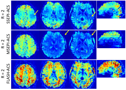
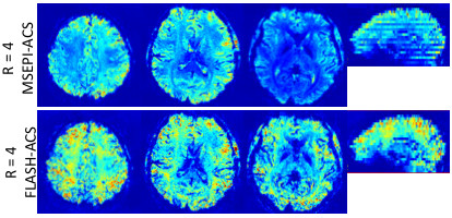
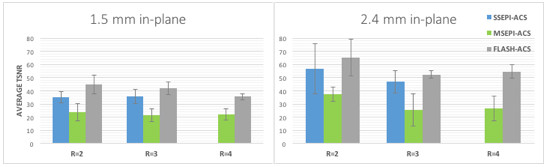
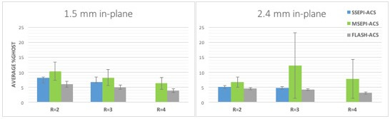
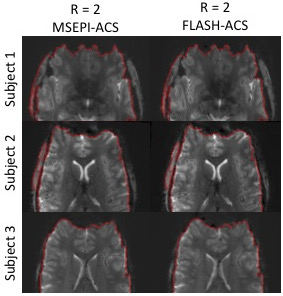
Figures 1 and 2 show representative tSNR maps for 1.5 mm in-plane resolution data from the same subject. Images reconstructed with FLASH-ACS have higher and more uniform tSNR throughout the brain compared to those obtained using SSEPI-ACS and MSEPI-ACS. SSEPI and MSEPI-ACS reconstructions exhibit heterogeneous tSNR within and across slices (Fig. 1&2 arrows and sagittal reformats). Figure 3A shows the comparison of the tSNR averaged across the whole brain, for all subjects. FLASH-ACS reconstructions are seen to produce significantly higher tSNR for all PAT factors and resolutions compared to SSEPI-ACS and MSEPI-ACS, with improvement from 60-100% over MSEPI-ACS reconstructions. Figure 4 shows graphs comparing the residual ghost level, averaged across all subjects. FLASH-ACS reconstructions have lower ghost level than SSEPI-ACS and MSEPI-ACS for all PAT factors and resolutions. Figure 5 shows example images with the most severe distortions, from three subjects, along with contour lines at the edge of the brain. Data show that edge contours are essentially identical for MSEPI-ACS and FLASH-ACS reconstructions in all cases.Discussion
In this study, we have demonstrated that the tSNR of GRAPPA accelerated EPI acquired at 7T can be improved significantly by using FLASH-ACS data during image reconstruction. An improvement in tSNR was seen for all PAT factors (R=2, 3, 4) and resolutions (1.5 mm and 2.4 mm). Reduction of tSNR when using SSEPI-ACS is attributed to phase errors in the ACS data that propagate into the GRAPPA kernel calculation (1). Because of the absence of phase errors, FLASH-ACS data provides a more accurate GRAPPA kernel and thus improved tSNR in reconstructed images. In addition, it was seen that the FLASH-ACS reconstructions have less residual EPI ghost and essentially identical image distortion characteristics compared to SSEPI-ACS and MSEPI-ACS. Therefore, use of the FLASH-ACS approach should directly benefit 7T fMRI studies using similar protocols, without adverse effects on image quality.Acknowledgements
No acknowledgement found.References
1) Talagala et al., MRM 75:2362 (2016)
2) Polimeni et al., MRM 75:665 (2016)
Figures