5161
Feature Refinement Scheme for Improved STEP Parallel Imaging Reconstruction1Philips Research China, Shanghai, People's Republic of China, 2Vascular Imaging Lab, Department of Radiology, University of Washington, WA, United States, 3Center for Biomedical Imaging Research, Department of Biomedical Engineering, Tsinghua University, Beijing, People's Republic of China
Synopsis
In this work, we incorporated the Feature Refinement (FR) scheme into the Self-supporting Tailored k-space Estimation for Parallel imaging reconstruction (STEP) image reconstruction framework to enhance its capability for structural representation in image domain, and developed a novel Weber Local Descriptor (WLD) method to improve the extraction of local image boundaries. With the preliminary experiments, it has been demonstrated that the improved STEP with WLD FR scheme can provide more accurate estimation of image details in comparison to original STEP and existing classic method.
Introduction
Parallel Imaging (PI)1,2 can enable fast magnetic resonance scans by exploiting the data redundancy across multiple channels, but the PI reconstruction method might cause image detail distortion due to aliasing artifact and noise amplification particularly in high acceleration factor condition. Previously, the Self-supporting Tailored k-space Estimation for Parallel imaging reconstruction (STEP) method3 has demonstrated that the spatially variant low rank signal model in k-space can improve the preservation of detail structure. However, the image detail structure is difficult to be directly extracted and represented in k-space domain. Recent study4 shows that iterative Feature Refinement (FR) in image domain can effectively capture the structural information from the residuals between pre- and post- compressed sensing filtered images within each iteration, but its extension for multi-channel PI method is nontrivial and the optimization of local image descriptor for structural information extraction is also required.Aim
To incorporate the FR scheme into the STEP image reconstruction framework and optimize the local image descriptor to further improve the reconstruction accuracy for image detail information.Methods
Incorporation of feature refinement
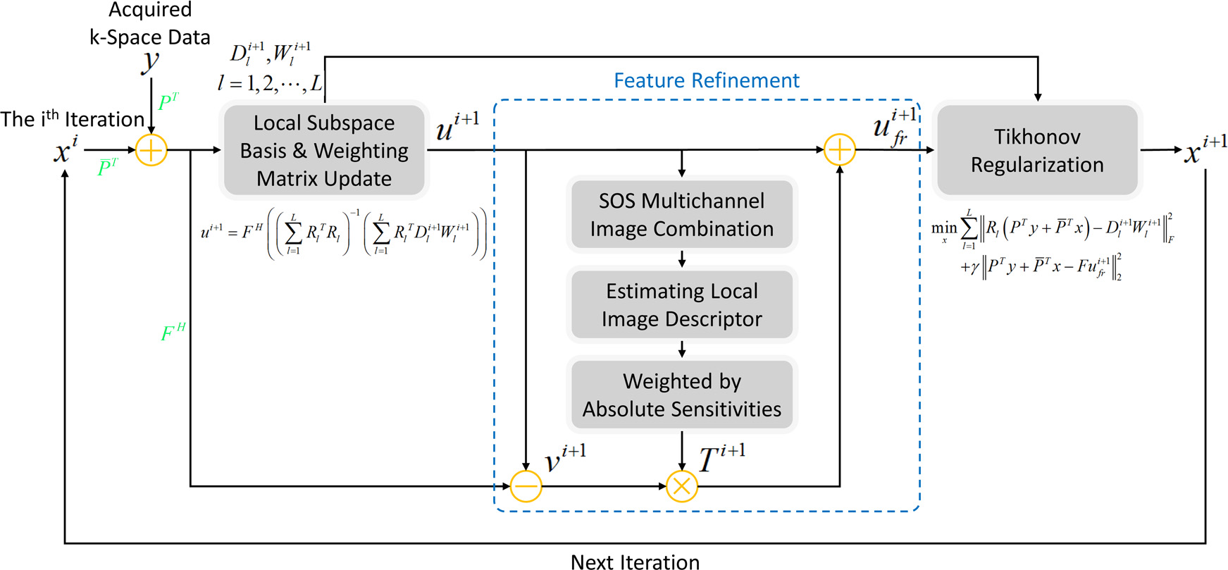
The FR module is introduced after the low rank and sparse approximation of local subspace basis and weighting matrix3. Since this approximation might suppress both noise-like artifacts and image details, it has the potential to capture some useful image features from the residuals and this feature refined image will be exploited as a Tikhonov regularization for the estimation of unacquired k-space data (Figure 1). The local image descriptor will be estimated on the Sum Of Squares (SOS) of the multi-channel images and weighted by the magnitude of sensitivity maps (which can be estimated by either ESPIRiT5 or scaling the magnitude image of each channel by the SOS image) to obtain its multi-channel counterpart, which can reduce the adverse impact of spatially varying signal-to-noise variations of surface coil for multi-channel image feature extraction.
Optimization of local image descriptor
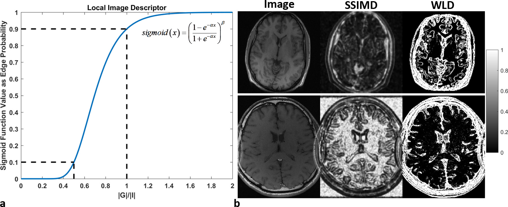
Inspired by the Weber’s law6, a Weber Local Descriptor (WLD) is introduced by establishing a nonlinear transform of the ratio between image gradient magnitude and intensity. The gradient magnitude is the SOS combination of multi-directional edge detection results (e.g. image convolution with Sobel operator) and dividing it by the image intensity can measure whether the local intensity variations are noticeable in perception. In addition, the nonlinear transform (e.g. sigmoid function) of this ratio (Figure 2a) can further highlight the location of edge boundaries while reduce the interference from slow variation of image contrast in comparison to the previously proposed Structural SIMilarity based Descriptor (SSIMD) (4) (Figure 2b). Since the image sharpness depends more on the edge preservation, it’s expected that the proposed WLD can more effectively enhance the image feature extraction.
Experiment setup
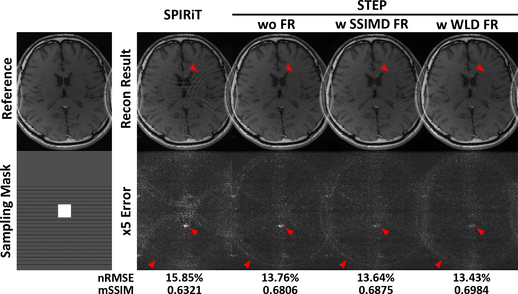
Two 8-channel Cartesian T1 weighted brain datasets with/without aliasing due to small field of view were obtained from the ESPIRiT toolbox on author’s webpage7, to evaluate the performance of improved STEP with FR scheme in comparison to SPIRiT and the original STEP method. Two typically used two dimensional undersampling patterns with central autocalibration signals (ACSs) were investigated on each dataset: variable density random undersampling (the size of ACSs: 26x26, acceleration factor (AF): x5) and uniform undersampling (the size of ACSs: 35x35, AF: x4). For a better assessment of FR effect, the subspace rank value and the basis selection threshold were fixed the same for STEP with/without FR scheme. The normalized Root Mean Square Error (nRMSE) and mean SSIM index (mSSIM)8 were exploited as two criterions for quantitative image quality measurement.
Results
In variable density random undersampling experiment, STEP can significantly reduce the noise amplification in comparison to SPIRiT, but it still fails to reduce the estimation errors on sharp image boundaries. The improved STEP with FR scheme can further alleviate these estimation errors particularly with the WLD FR scheme. The comparison of local zoom-in views from different reconstruction methods can also demonstrate the improved image detail preservation of the improved STEP with WLD FR. Similarly in uniform undersampling experiment, the edge information can be better estimated by the improved STEP with FR scheme. In addition, the aliasing artifacts can be also further suppressed in the improved STEP with WLD FR.Discussion and Conclusion
In this work, we incorporated the FR scheme into the STEP image reconstruction framework to enhance its structural representation in image domain, and proposed a novel WLD method to improve the extraction of local image boundaries. With the preliminary results, it has been demonstrated that the improved STEP with WLD FR scheme can provide more accurate estimation of image details.Acknowledgements
No acknowledgement found.References
1. Pruessmann KP, Weiger M, Scheidegger MB, et al. SENSE: sensitivity encoding for fast MRI. Magn Reson Med. 1999;42(5):952-962.
2. Griswold MA, Jakob PM, Heidemann RM, et al. Generalized autocalibrating partially parallel acquisitions (GRAPPA). Magn Reson Med. 2002;47(6):1202-1210.
3. Zhou Z, Wang J, Balu N, et al. STEP: Self-supporting tailored k-space estimation for parallel imaging reconstruction. Magn Reson Med. 2016;75(2):750-761.
4. Wang S, Liu J, Liu Q, et al. Iterative feature refinement for accurate undersampled MR image reconstruction. Phys Med Biol. 2016;61(9):3291-3316.
5. Uecker M, Lai P, Murphy MJ, et al. ESPIRiT--an eigenvalue approach to autocalibrating parallel MRI: where SENSE meets GRAPPA. Magn Reson Med. 2014;71(3):990-1001.
6. Chen J, Shan S, He C, et al. WLD: A Robust Local Image Descriptor. IEEE Trans Pattern Anal Mach Intell. 2010;32(9):1705-1720.
7. Lustig M, Pauly JM. SPIRiT: Iterative self-consistent parallel imaging reconstruction from arbitrary k-space. Magn Reson Med. 2010;64(2):457-471.
8. Wang Z, Bovik AC, Sheikh HR, et al. Image quality assessment: from error visibility to structural similarity. IEEE Trans Image Process. 2004;13(4):600-612.
Figures