5155
T2-Mapping Relaxation Time Correlates with Histopathological Degree of Degeneration in Meniscal Tissue1Radiology and Nuclear Medicine, Erasmus MC, Rotterdam, Netherlands, 2Orthopedic Surgery, Erasmus MC, Rotterdam, Netherlands, 3Medical Informatics, Erasmus MC, Rotterdam, Netherlands
Synopsis
To validate meniscal T2-mapping, which is suggested to detect early meniscal degeneration, T2 relaxation times of ten menisci (five traumatically torn and five degenerative menisci from 10 patients) were compared to histopathology as the gold standard to assess meniscal degeneration. T2 relaxation times were found to correlate well with histopathological degree of degeneration in meniscal tissue (rs = 0.64, P = 0.001). Degenerative meniscal tissue was found to exhibit significantly higher T2 relaxation times compared to non-degenerated meniscal tissue (22.7±3.0 ms and 18.2 ±5.1 ms (P = 0.02) respectively). Therefore, T2-mapping is a promising quantitative imaging biomarker for meniscal degeneration.
INTRODUCTION
To optimize treatment selection for meniscal pathologies and to improve patient monitoring, a sensitive non-invasive imaging technique for the meniscus is needed. T2-mapping is a promising quantitative MRI technique that is suggested to detect early meniscal degeneration. However, very few studies have validated meniscal T2-mapping against histopathological findings as the gold standard, and studies investigating non-degenerated meniscal tissue are lacking completely. Therefore, our aim was to study the correlation between T2-mapping relaxation times and the degree of histopathological degeneration in both degenerated- and non-degenerated meniscal tissue.METHODS
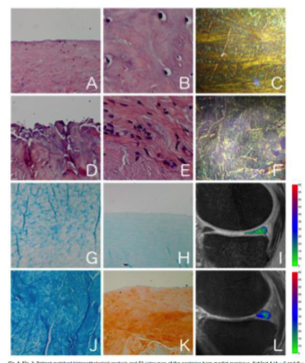
In this prospective observational study 10 patients were included, both with degenerative and non-degenerative menisci. Traumatically torn meniscal tissue from five patients was collected during arthroscopic partial meniscectomy. Degenerative menisci from five patients were obtained during total knee replacement surgery. All patients underwent a pre-operative MRI scan at 3 Tesla which included a 3D FSE T2-mapping pulse sequence with 5 echo times1. Histopathological analysis was performed using the Pauli score2, a validated semi-quantitative grading system involving the following subdomains: surface integrity, cellularity, matrix and collagen organisation, and proteoglycan staining intensity (Fig. 1 A-H, J, K). Mean T2 relaxation times were calculated in meniscal regions of interest corresponding with the areas scored histopathologically, using in-house developed post-processing software (Fig 1 I and L). The correlation between T2-mapping and histopathological degeneration was assessed using Spearman correlation tests.RESULTS
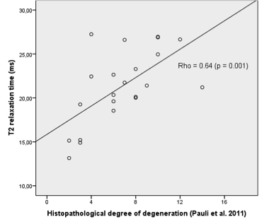
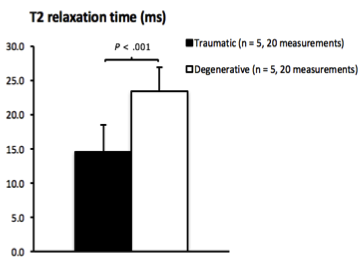
Meniscus T2 relaxation time correlated well with the degree of histopathological degeneration (rs = 0.64, P = 0.001, Fig 2). Meniscal tissue with mild to moderate degeneration was found to exhibit statistically significantly higher T2 relaxation times compared to meniscal tissue without degeneration: 22.7 ± 3.0 ms and 18.2 ± 5.1 ms (P = 0.02), respectively (Fig 3).DISCUSSION
Despite our relatively small sample size, we found a highly significant correlation between T2-mapping relaxation time and the degree of histopathological degeneration in meniscal tissue. These results warrant further research in larger patient samples and with the use of other T2-mapping acquisition methods.CONCLUSION
T2-mapping relaxation time correlates well with histopathological degeneration in meniscal tissue and is a promising quantitative imaging biomarker to estimate meniscal degeneration.Acknowledgements
No acknowledgement found.References
1. Chen et al. 19th edition of the ISMRM: Annual Meeting & Exhibition. Montréal; 2011
2. Pauli C, Grogan SP, Patil S, Otsuki S, Hasegawa A, Koziol J, Lotz MK, D'Lima DD. Macroscopic and histopathologic analysis of human knee menisci in aging and osteoarthritis. Osteoarthritis Cartilage. 2011 Sep;19(9):1132-41
Figures