5152
Dynamic Contrast-Enhanced Magnetic Resonance Imaging Parameters in Metastatic Bone Lesions of Non-small Cell Lung Cancer: Comparison between Lesions with and without Epidermal Growth Factor Receptor (EGFR) Mutation in Primary Cancer1Radiology, Samsung medical center, Seoul, Korea, Republic of
Synopsis
The presence of the epidermal growth factor receptor (EGFR) gene mutation is associated with a high rate of distant metastasis and poor prognosis in patients with non-small cell lung cancer (NSCLC). EGFR signaling acts as mediator of bone metastasis by increasing tumor cell proliferation and promotes synthesis and secretion of numerous angiogenic growth factors. We propose that metastatic bone lesions with and without the EGFR mutation in primary NSCLC have different perfusion profiles and dynamic contrast-enhanced magnetic resonance imaging (DCE-MRI) parameters can reveal the differences.
Purpose
To compare dynamic contrast-enhanced magnetic resonance imaging (DCE-MRI) parameters between metastatic bone lesions with and without the epidermal growth factor receptor (EGFR) gene mutation in consecutive patients with primary non-small cell lung cancer (NSCLC).Materials and methods
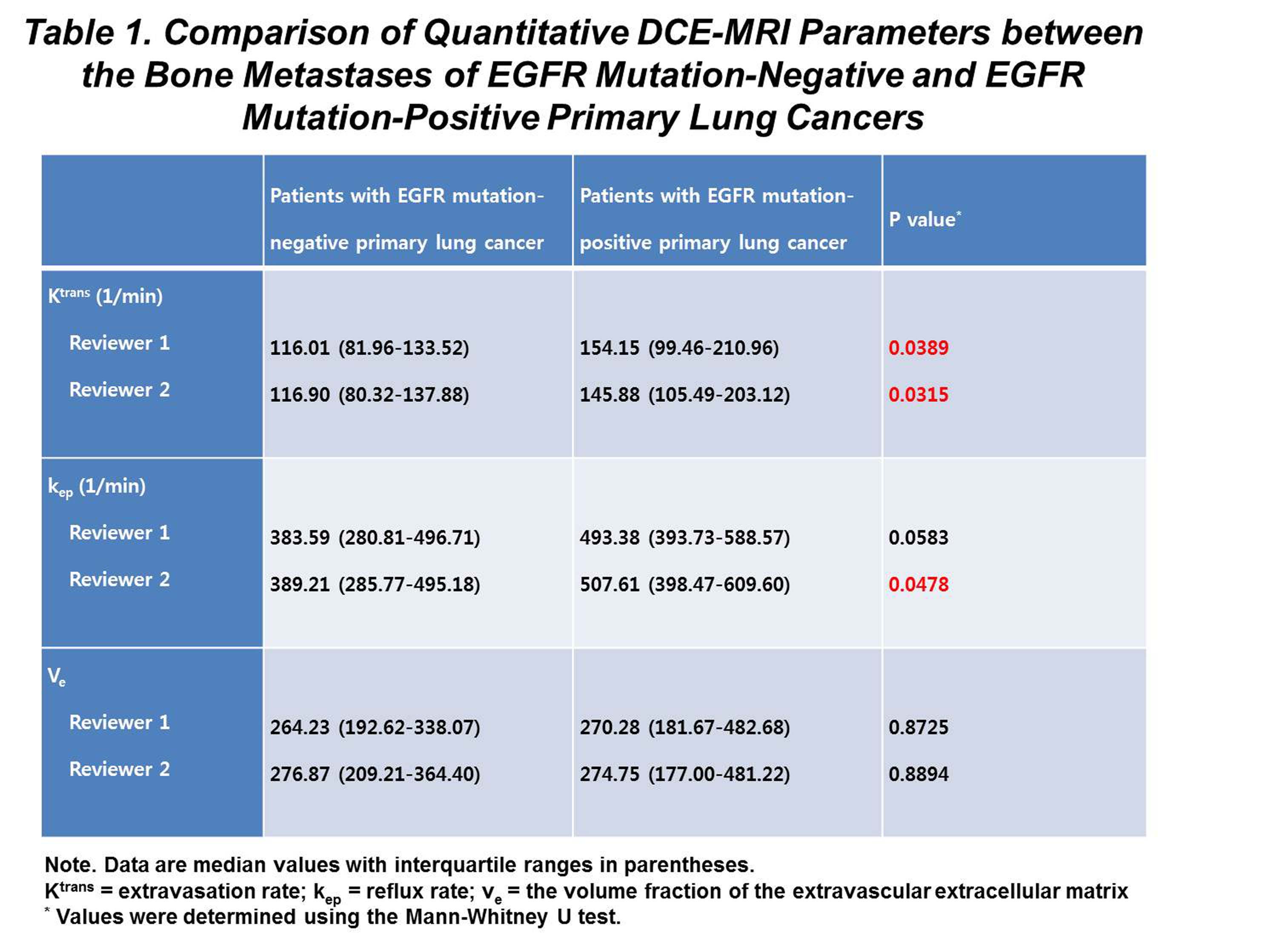
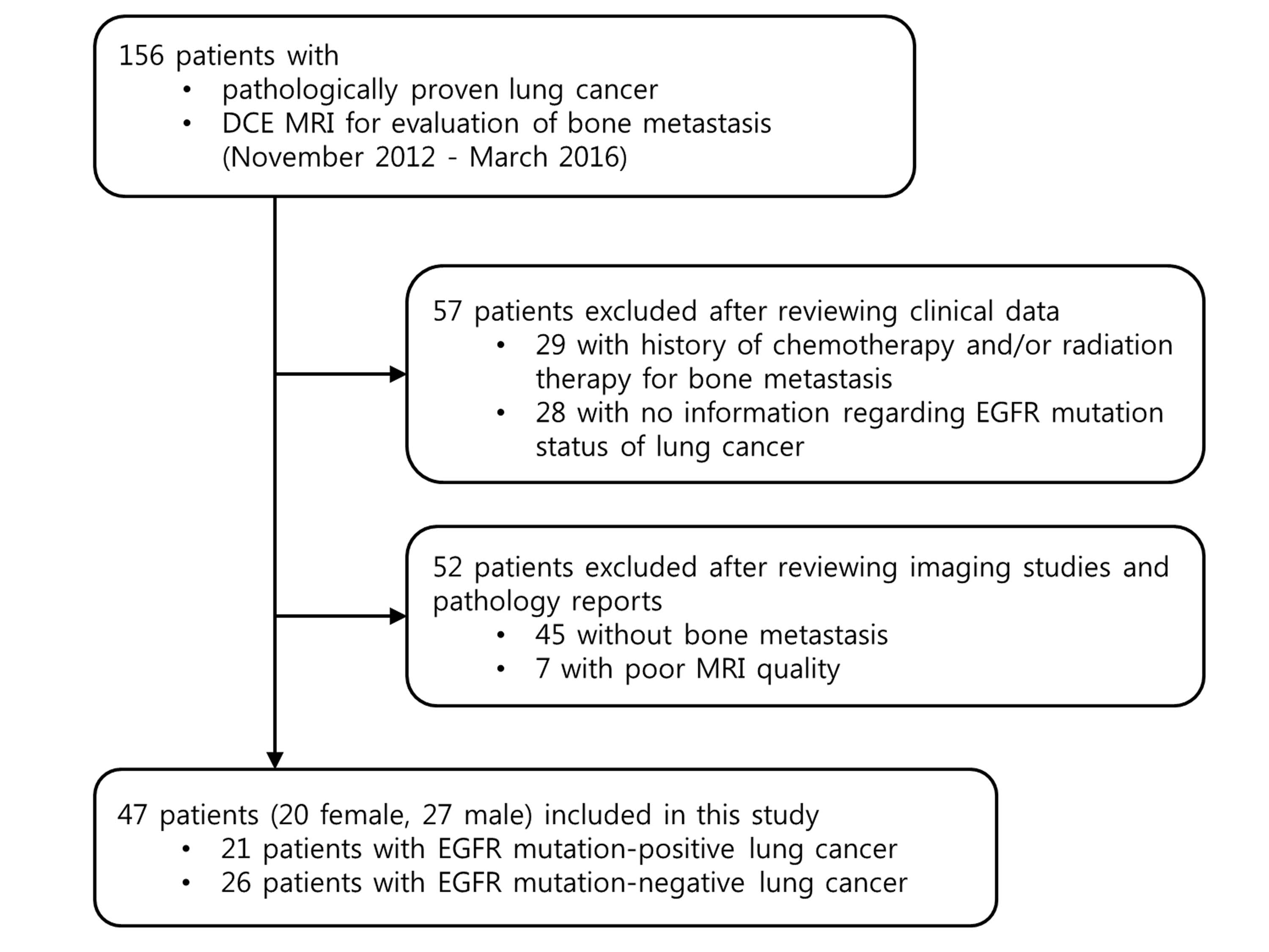
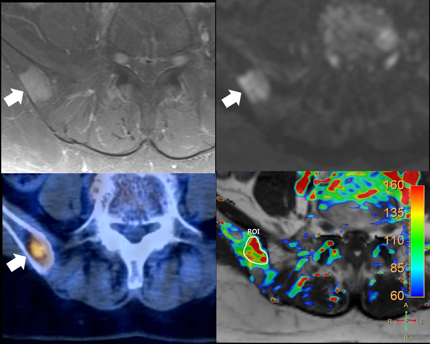
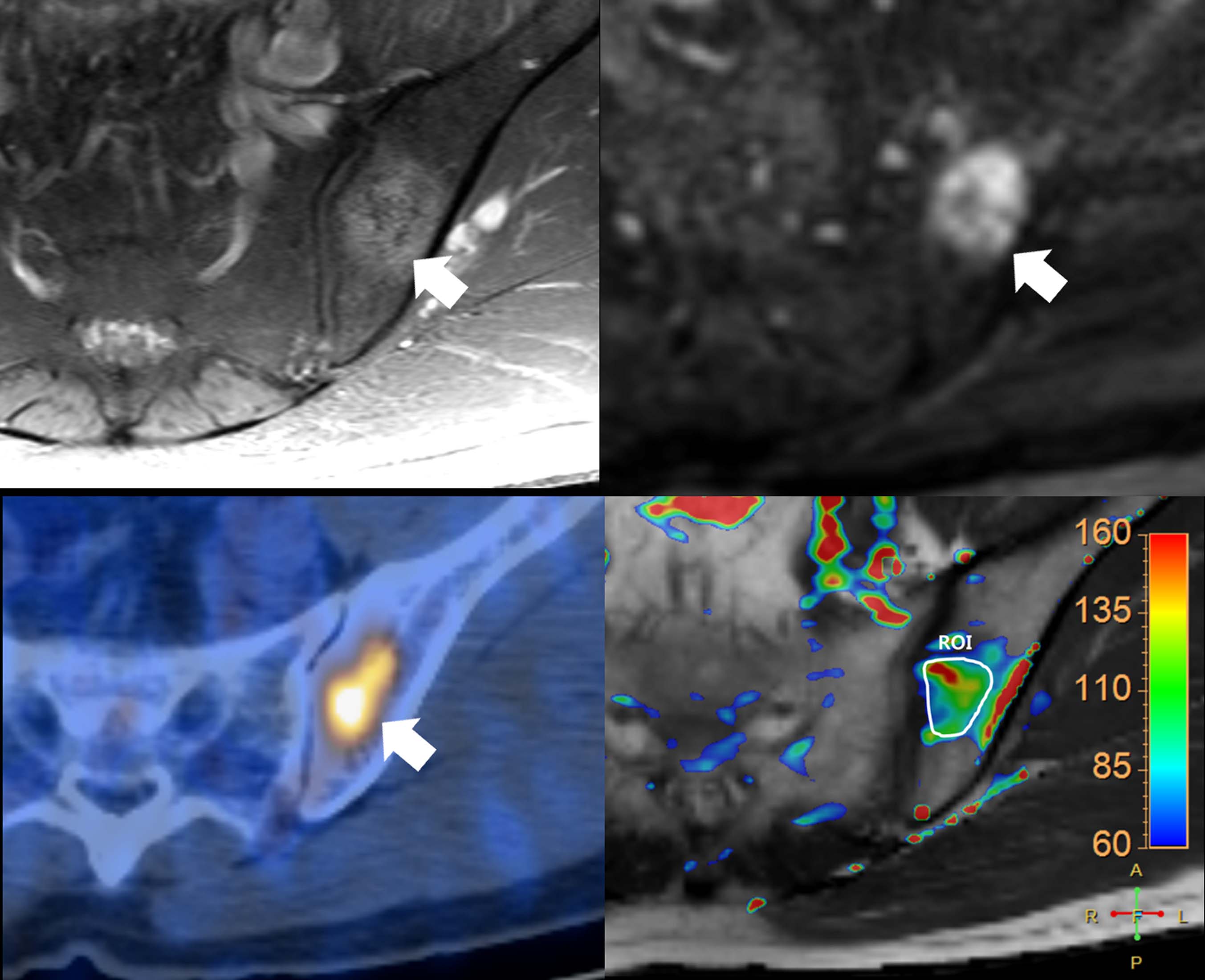
Forty-seven patients with NSCLC and a confirmed EGFR mutation status (21, EGFR mutation-positive; 26, EGFR mutation-negative), who underwent DCE-MRI for bone metastases between November 2012 and March 2016, were included in this IRB-approved study. Two independent radiologists performed analysis of extravasation rate (Ktrans), reflux rate (kep), and the volume fraction of the extravascular extracellular matrix (ve) using image-processing software. Intergroup comparisons of the mean measured parameters were performed using the Mann-Whitney U test. Interobserver agreement was calculated using the intraclass correlation coefficient.Results
Analysis revealed a high level of agreement between the two reviewers for all three parameters (0.95 for Ktrans, 0.97 for kep, and 0.91 for ve). Ktrans was significantly higher in the EGFR mutation-positive group (P = 0.0389 for reviewer 1; 0.0315 for reviewer 2). kep was also higher in the EGFR mutation-positive group, but showed statistical significance only in the evaluation performed by one reviewer (P = 0.0478 for reviewer 2; 0.0583 for reviewer 1). No significant difference was observed in ve (P = 0.8725 for reviewer 1; 0.8894 for reviewer 2).Conclusion
Our study result has shown a significantly higher value of Ktrans in metastatic bone lesions of EGFR mutation-positive primary lung cancer than in those of EGFR mutation-negative primary lung cancer. A similar tendency was demonstrated for kep on intergroup comparisons. These results may reflect the histologic characteristic of bone metastasis from EGFR mutation-positive lung cancer related to an increased angiogenic activity. The findings may also form the basis for the possible use of DCE-MRI parameters for evaluating treatment response in these lesions in future studies.Acknowledgements
No acknowledgement found.References
1. Togashi Y, Masago K, Kubo T, et al. Association of diffuse, random pulmonary metastases, including miliary metastases, with epidermal growth factor receptor mutations in lung adenocarcinoma. Cancer 2011;117(4):819-825.
2. Fujimoto D, Ueda H, Shimizu R, et al. Features and prognostic impact of distant metastasis in patients with stage IV lung adenocarcinoma harboring EGFR mutations: importance of bone metastasis. Clin Exp Metastasis 2014;31(5):543-551.
3. Lu X, Kang Y. Epidermal growth factor signalling and bone metastasis. Br J Cancer 2010;102(3):457-461.
4. De Luca A, Carotenuto A, Rachiglio A, et al. The role of the EGFR signaling in tumor microenvironment. J Cell Physiol 2008;214(3):559-567.
Figures