5151
Feasibility study of intra-voxel incoherent motion MR imaging for the differentiation of benign and malignant bone tumors1Zhengzhou University First Affiliated Hospital, Zhengzhou,Henan, People's Republic of China, 2Clinical Science, Philips Healthcare, People's Republic of China
Synopsis
This work investigated and evaluated the role of intra-voxel incoherent motion(IVIM) MR imaging in characterizing the bone tumors, and furtherly evaluate the ability of IVIM parameters to differentiate benign and malignant tumors by using a support vector machine (SVM) classifier, which might be helpful for clinical diagnosis and studies.
Purpose
Recent studies have shown that IVIM offers a method to reflect the information of perfusion and diffusion in complex biological tissues in various brain diseases1,liver diseases2, and breast tumors3, but rarely used in the study of bone tumors.The purpose of this study is to investigate the application of IVIM on bone tumors and furtherly evaluate the ability of IVIM parameters to differentiate benign and malignant tumors by using a support vector machine (SVM) classifier.Methods
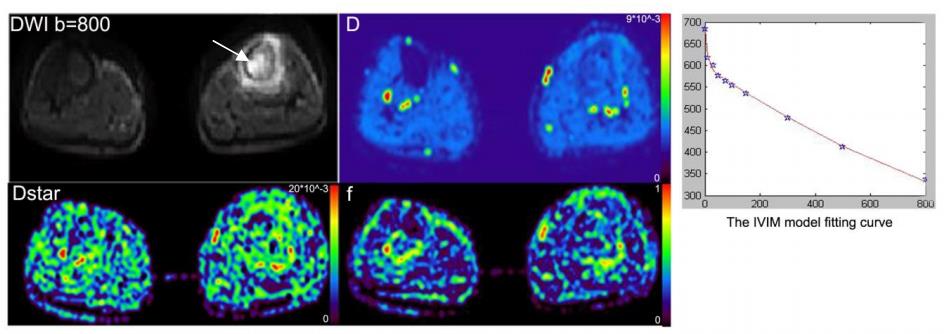
Thirty-one patients (15 males and 16 females aged 37.9±18.2 years old) with bone tumors (diagnosed according to pathological biopsy) were included in this study.Based on the WHO Classification of Tumors of Soft Tissue and Bone (2012) criteria, 31 patients were divided into two groups:11 for benign tumors and 20 for malignancies. All the patients were scanned by MR IVIM sequence based on a 3T MR scanner (Ingenia, Philips Healthcare, Best, the Netherlands). The IVIM scanning was performed with 10 b-values of 0, 10, 30, 50, 75, 100, 150, 300, 500, 800s/mm2.The IVIM related parameters of tissue diffusivity (D), pseudo-diffusion coefficient (D* or Dstar) and perfusion fraction (f) were calculated by using a Philips development software (Version 3.0). All the above parameters were measured by drawing ROIs (Region of Interest) within the periphery of the lesions(P_D,P_Dstar,P_f short for periphery D, periphery Dstar,periphery f),the center of the lesions(C_D,C_Dstar,C_f short for center D, center Dstar, center f), the normal bone marrow(NB_D,NB_Dstar,NB_f short for normal bone marrow D, normal bone marrow Dstar, normal bone marrow f,) and the normal soft tissue(NS_D,NS_Dstar,NS_f short for normal soft tissue D, normal soft tissue Dstar, normal soft tissue f,). Besides, some new parameters (such as P_NB_Dstar means the subtraction of NB_Dstar from P_Dstar, and P_NB_NB_Dstar means the NB_Dstar divided by the P_NB_Dstar) were calculated. And the all the parameters extracted from benign and malignant patients were compared by using Mann-Whitney U test with SPSS software (version 16.0). A P value of less than 0.05 was considered statistically significant. And receiver operating characteristic (ROC) analysis was performed to assess the sensitivity and specificity of every parameter in the diagnosis of benign and malignant bone lesions.The parameters which showed a significant difference between benign and malignant lesions were selected and combined as the feature vectors for the furtherly classification by using SVM classifier.Results
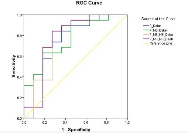
The results revealed that P_Dstar value in the lesions is significantly different (P <0.05), moreover the values of P_NB_Dstar, P_NB_NB_Dstar and P_NS_NS_Dstar also showed significant difference (P<0.05)between benign and malignant lesions.The ROC analysis results were shown in Figure 2,which showed the ability of the above parameters to differentiate benign and malignant lesions.And the SVM classifier showed a high classification accuracy (80.65%), sensitivity (85%) and specificity (72.73%) for differentiating malignant from the benign lesions.Discussion
IVIM is a non-invasive functional imaging based on diffusion MRI technique, which provide simultaneous evaluation of tumor vascularization and diffusion for the bone tumor characterization. According to IVIM principle, Dstar value and f value are perfusion related parameters, while D value reflect the real water molecule motion.The application of IVIM in other systems has been reported a lot previously, IVIM related parameters shows a good diagnostic efficiency1, and perfusion parameters reflect structure of the microvascular in the tumor. Dstar maps has conformity with the known histologic diagnosis2.Our study results show that P_Dstar and related parameters are able to differentiate benign from malignant bone tumors, which suggest that D* value provide vascularize information of tumor. Meanwhile, in this study, the SVM showed a good performance in the differentiation of malignant tumors from benign ones and this would assist doctors to diagnose the type of the tumor and furtherly was helpful for the corresponding treatment plan for the tumor. In the future, more patients with different types of bone tumors will be included in our study to evaluate clinical value of the IVIM technique and other types of classifiers.Conclusions
IVIM technique is helpful to evaluate the pathological behavior and provide useful information on the perfusion and diffusion properties related to bone tumors microenvironment. Different Dstar related parameters increases diagnostic confidence of bone tumors.Acknowledgements
No acknowledgement found.References
1.VincenzaGranata, Roberta Fusco, Orlando Catalano, et al. Intra-voxel incoherent motion (IVIM) in diffusion-weighted imaging (DWI) for Hepatocellular carcinoma: correlation with histologic grade [J].Oncotarget, 2016,[Epub ahead of print].
2. C. Federau, R. Meuli, K. O’Brien, et al. Perfusion Measurement in brain gliomas with intra-voxel incoherent motion MRI [J].Am J Neuroradiol,2014,35: 256-262.
3. Jing Yuan1, Oi Lei Wong, Gladys G. Lo, et al. Statistical assessment of bi-exponential diffusion weighted imaging signal characteristics induced by intra-voxel incoherent motion in malignant breast tumors[J]. Quant Imaging Med Surg, 2016,6(4):418-429.
Figures