5150
Feasibility study of diffusional kurtosis imaging for the differentiation of musculoskeletal benign and malignant tumors1Zhengzhou University First Affiliated Hospital, Zhengzhou,Henan, People's Republic of China, 2Clinical Science, Philips Healthcare, People's Republic of China
Synopsis
This work investigated and evaluated the role of magnetic resonance (MR) diffusion kurtosis imaging(DKI) in characterizing the musculoskeletal tumors, and furtherly evaluate the ability of DKI parameters to differentiate benign and malignant tumors by using a classifier based on random forest method, which might be helpful for clinical diagnosis and studies.
Purpose
Recent studies have shown that DKI provides a new method to evaluate the non-gaussian diffusion behavior in complex biological tissues in various brain diseases1and breast tumors2, but it was rarely used in the study of musculoskeletal tumors.The purpose of this study is to investigate the application of DKI on musculoskeletal tumors and furtherly evaluate the ability of DKI parameters to differentiate benign and malignant tumors by using a classifier based on random forest method.Methods
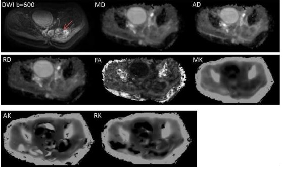
Twenty-eight patients (15 males and 13 females aged 37±1.9 years old) with musculoskeletal tumors (all have been diagnosed as musculoskeletal tumor according to pathological biopsy)were included in this study.And based on the WHO Classification of Tumors of Soft Tissue and Bone(2012) criteria, 28 patients were divided into two groups:10 for benign tumors and 18 for malignancies. All the patients were scanned by MR DKI sequence based on a 3T MR scanner (Ingenia, Philips Healthcare, Best, the Netherlands). The DKI scanning was performed with 3 b-values of 0, 600, 1,200 s/mm2 and 15 motion-sensitive gradient directions.The DTI related parameters of mean diffusivity (MD),fractional anisotropy(FA), axial diffusivity (AD), radical diffusivity (RD) and DKI related parameters of mean kurtosis (MK), axial kurtosis(AK), radical kurtosis(RK) were calculated by using DKE software (Version 2.6.0, website:www.musc.edu/cbi). All the above parameters were measured by drawing ROIs (Region of Interest) within the periphery and center of the lesions. And the measured parameters in benign and malignant lesions were compared by using Mann-Whitney U test with SPSS software (version 16.0). A P value of less than 0.05 was considered statistically significant. And receiver operating characteristic (ROC) analysis was performed to assess the sensitivity and specificity of every parameter in the diagnosis of benign and malignant musculoskeletal lesions.The parameters which showed a significant difference between benign and malignant lesions were selected and combined as the feature vectors for the furtherly classification by using random forest classifier. Results
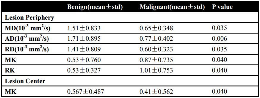
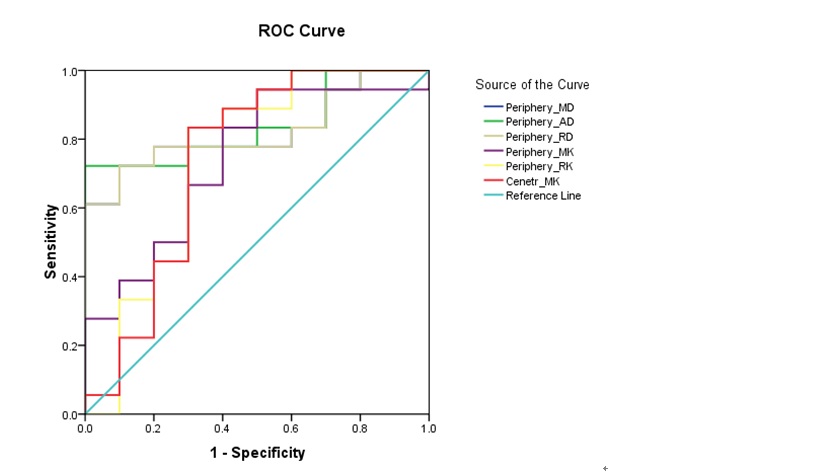
The statistical results of the parameters with significant difference between benign and malignant lesions were summarized in Table 1. The results revealed that MD, AD, RD, MK,RK values of the periphery and MK value of the center in the lesions are significantly different (P <0.05). The ROC analysis results were shown in Figure 2,which showed the ability of MD, AD, RD, MK,RK values of the lesion periphery and MK value of the lesion center to differentiate benign and malignant lesions. And the classifier showed a high classification accuracy (85.7%), sensitivity (88.9%) and specificity (80%) for differentiating malignant from the benign lesions.Discussion
DKI is a non-invasive functional imaging based on diffusion MRI technique, which provide useful information of tumor cytoarchitectonic complexity on the water diffusion properties3.Our study results show that the DTI and DKI related parameters is able to differentiate benign from malignant musculoskeletal tumors. Meanwhile, according to our analysis, MK value has the ability of characterizing musculoskeletal tumors, which reflect microstructure differences between benign and malignant tumors. Random forest is effective in the classification of datasets in many fields such as sociology and medical science. In this work, the random forest showed a better performance in the differentiation of malignant tumors from benign ones and this would assist doctors to diagnose the type of the tumor and furtherly was helpful for the corresponding treatment plan for the tumor. In the future, more patients with different types of musculoskeletal tumors will be included in our study to evaluate clinical value of the DKI technique and other types of classifiers.Conclusions
DKI technique is helpful to evaluate the pathological behavior and provide useful information on the diffusion properties related to musculoskeletal tumors microenvironment. The multivariate pattern analysis of DKI parameters increases diagnostic confidence of musculoskeletal tumors.Acknowledgements
No acknowledgement found.References
1. Peter Raab, Elke Hattingen ,Kea Franz, et al.Cerebral Gliomas: Diffusional Kurtosis Imaging Analysis of Microstructural Differences[J].Radiology,2010,254(3):876-881.
2. LuísaNogueira, Sofia Brandão, Eduarda Matos,et al.Application of the diffusion kurtosis model for the study of breast lesions[J].Eur Radiol,2014,24:1197-1203.
3.Dongmei Wu, Guanwu Li, Junxiang Zhang,et al.Characterization of Breast Tumors Using Diffusion Kurtosis Imaging (DKI)[J].PLOS ONE,2014,9(11):e113240.
Figures