5149
Characterization of benign and malignant soft tissue tumors using dynamic contrast enhanced magnetic resonance imagingYing Li1, Cuiping Ren1, Jingliang Cheng1, and Zhizheng Zhuo2
1Zhengzhou University First Affiliated Hospital, Zhengzhou,Henan, People's Republic of China, 2Clinical Science, Philips Healthcare, People's Republic of China
Synopsis
This work investigated and evaluated the role of dynamic contrast enhanced magnetic resonance imaging(DCE-MRI) in characterizing the soft tissue tumors, and furtherly evaluate the ability of permeability parameters to differentiate benign and malignant tumors by using random tree and artificial neural network classifiers, which might be helpful for clinical diagnosis and studies.
Purpose
Recent studies have shown that dynamic contrast enhanced magnetic resonance imaging(DCE-MRI) especially permeability offers a method to reflect the information of vascular permeability and density of micro-vesselsin tumors1,2, but rarely used in the study of soft tissue tumors.The purpose of this study is to investigate the clinical application of DCE imaging on soft tissue tumors and furtherly evaluate the ability of the related permeability parameters to differentiate benign and malignant tumors by using random tree and artificial neural network classifiers.Methods
Twenty-one patients (8males and 13 females aged 38.6±19.3 years old) with soft tissue tumors (all have been diagnosis as soft tissue tumor according to pathological biopsy) were included in this study.And based on the WHO Classification of Tumors of Soft Tissue and Bone(2012) criteria, 21 patients were divided into two groups:13 for benign tumors and 8 for malignancies. Each subject underwent a MR scanning by using a 3T MR scanner (Ingenia, Philips Healthcare, Best, the Netherlands).All the patients were scanned by permeability (time-resolved 3D TFE) sequence with following parameters:TR/TE=4.5ms/2.1ms, FA=10 degree,voxel size=2mm×2mm×2mm,various field of view and matrix size according to the anatomy size, 50 dynamics within scan time of 4min-6min, and multiple FA of 5,10 and 15 degrees before the Gadolinium contrast agent injection for calculating T1 maps. Toft’s model was used for calculation of Ktrans (volume transfer constant),Kep (microvascular permeability reflux constant),Ve(extra-vascular extracellular space distribute volume per unit tissue volume),Vp (fractional plasma volume) and AUC(area under curve).All the above parameters were measured by drawing ROIs (Region of Interest) within the periphery of the lesions(P_Ktrans,P_Kep,P_Ve ,P_Vp,P_AUC short for periphery Ktrans, periphery Kep, peripheryVe, peripheryVp, peripheryAUC) and the center of the lesions(C_Ktrans, C_Kep,C_Ve ,C_Vp short for center Ktrans, center Kep,center Ve, center Vp, center AUC). Besides, some new parameters (such as P_C_Ktrans means the subtraction of C_Ktrans from P_Ktrans) were calculated. And the measured parameters in benign and malignant lesions were compared by using Mann-Whitney U test with SPSS software (version 16.0). A P value of less than 0.05 was considered statistically significant. And receiver operating characteristic (ROC) analysis was performed to assess the sensitivity and specificity of the parameter those showed significant differences between benign and malignant soft tissue lesions. The parameters which showed a significant difference between benign and malignant lesions were selected and combined as the feature vectors for the furtherly classification by random tree and artificial neural network classifiers.Results
The results revealed that Ktrans,Kep,Ve ,Vp and AUC values in the lesions is not significantly different (P>0.05). However, P_C_Ktrans,P_C_Vp and P_C_AUC values showed significant difference (P<0.05)between benign and malignant lesions.The ROC analysis results were shown in Figure 1,which showed the ability of the above parameters to differentiate benign and malignant lesions.And the random tree classifier showed a high classification accuracy (81%), sensitivity (75%) and specificity (84.6%) for differentiating malignant from the benign lesions (as shown in Table 1).Discussion
DCE related parameters provide the information of vascular permeability and density of micro-vessels in characterizing the soft tissue tumors, which can reflect the kind of the mass and further the malignant degree. Massive pathological micro-vessels around bone tumor would generate, which have an incomplete structure with a high permeability. Moreover, the periphery of the lesion is rich in vascular endothelial growth factor which leads to abundant blood supply in the boundary, while the center of lesion is poor blood supply. The subtraction of permeability parameters within center region from those within periphery region has conformity with the known histologic diagnosis, which consistent with this study. Meanwhile, in this study, the random tree showed a good performance in the differentiation of malignant tumors from benign ones and this would assist doctors to diagnose the type of the tumor and furtherly was helpful for the corresponding treatment plan for the tumor.Conclusions
Permeability parameters can provide useful information on the vascular permeability and density of micro-vessels in characterizing the soft tissue tumors, especially the difference of periphery and center of the lesions increase the differentiation accuracy of the malignant from benign tumors.Acknowledgements
No acknowledgement found.References
1.Alonzai R, Padhani AR, Allen C. Dynamic contrast enhanced MRI in prostate cancer[J].Eur J Radiol,2007,63(3):335-350.
2.Pickles MD, Lowry M, Manton DJ, et al. Role of dynamic contrast enhanced MRI in monitoring early response of locally advanced breast cancer to neoadjuvant chemotherapy[J].Breast Cancer Res Treat,2005,91(1):1-10.
Figures
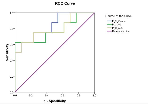
Figure 1. The ROC analysis
results of the P_C_Ktrans, P_C_Vp and P_C_AUC for the differentiation the
malignant from benign soft tissue tumors. The area under the ROC curve are
0.777, 0.746 and 0.838 respectively.
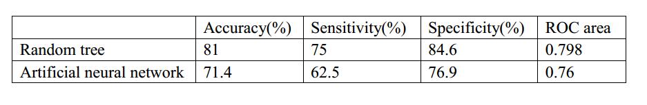
Table 1. The classification
results of random tree and artificial neural network for the differentiation
the malignant from benign soft tissue tumors.