5143
Improved visualization of diffusion-prepared MR neurography (SHINKEI) in the pelvis using high-intensity reduction (HIRE) techniqueHitoshi Tadenuma1, Kayoko Abe2, Masami Yoneyama3, Yasuhiro Goto1, Makoto Suzuki1, Mamoru Takeyama1, and Shuji Sakai2
1Department of Radiological Service, Tokyo Women’s Medical University, Tokyo, Japan, 2Department of Diagnostic Imaging and Nuclear Medicine, Tokyo Women’s Medical University, Tokyo, Japan, 3Philips Electronics Japan, Ltd, Tokyo, Japan
Synopsis
The role of MRI in the diagnosis of Lumbrosacral plexopathy is in depiction of the anatomical detail of pelvic nerves. This study attempted the improvement of the pelvic nervous depiction using HIRE-SHINKEI. This applies HIRE technique, and requires both SHINKEI and additional 3D heavily T2W scan to subtract the data. HIRE-SHINKEI can improve the depiction of nerves by subtracting signals of the background. This new technique has great potential to help the diagnosis for the peripheral nerve disorders of the lumbosacral region.
Background & Purpose
MRI has not made a substantial contribution in the diagnosis peripheral nerve disorders in the lumbosacral region because it was difficult to visualize the lumbosacral plexus clearly using ordinary MR techniques1-3. Recently, improved motion-sensitized driven-equilibrium (iMSDE) has been developed to suppress blood flow signals, and MR neurography of the lumbar plexus based on iMSDE technique was reported as a new method named nerve-SHeath signal increased with INKed rest-tissue RARE Imaging (SHINKEI)4,5. However, signal suppression of vein, bladder, and cerebrospinal fluid (CSF) was still not enough to visualize the peripheral side of the lumbosacral nerves on SHINKEI. High-intensity reduction technique (HIRE) was also reported as a technique which subtracts two images with different echo times to suppress signal intensity of the T2 elongated area6. Therefore, we assessed a new technique (HIRE-SHINKEI), a combination of SHINKEI and HIRE, to suppress signal intensities of CSF, bladder, and veins in the Pelvis, which leads to improvement of lumbosacral plexus detection.Method
HIRE is a subtraction technique. It obtains two images with different TE, on which signals of CSF, bladder, and veins are considerably attenuated (Fig. 1). SHINKEI based on 3D T2WI applies two specific pre-pulses; one of which is a fat-suppression pre-pulse, another of which is iMSDE pre-pulse to suppress signals from vessels. The peripheral nerves show high signal intensity on SHINKEI. TE value on 3D heavily T2W is set longer than on SHINKEI. On 3D heavily T2W, the peripheral nerves show low signal intensity, although signal intensities of CSF, bladder, and veins are as high as those on SHINKEI. Therefore, we applied HIRE-SHINKEI, on which images on 3D heavily T2W and SHINKEI were subtracted to suppress signals of CSF, bladder, and veins in expectation of improved demonstration of the lumbosacral plexus (Fig. 2). Six volunteers were examined with 3T MR unit (Ingenia 3T, Philips Healthcare R5.1.7) using a 32-element torso coil. The study was approved by the local institutional review board. The imaging parameters of SHINKEI and 3D heavily T2W were: TR = 2400 ms, FOV = 380*380 (mm), voxel size of 1.1*1.1*1 mm 3. SHINKEI imaging parameters were: Contrast Equivalent TE = 68 ms, TSE factor = 100, and total acquisition time = 5 m 52 s. 3D heavily T2W parameters were; TE depends on TSE factors, and total acquisition time was 3 m 48 s.Results & Discussion
Volunteer studies were successfully performed, resulting in clear depictions of pelvic nerves. Representative images of MR neurography of the pelvis by using HIRE-SHINKEI are shown in Fig. 3. Signals of CSF, bladder and veins were clearly suppressed in the pelvis. Compared to SHINKEI, pelvic splanchnic nerves were detected more clearly on HIRE-SHINKEI. Sciatic nerves were detected from the origin at sacral spinal nerves I to III. Pelvic splanchnic nerves and pudendal nerves were also detected clearly (Fig. 4). Long TE commonly leads to acquisition time extension on 3D heavily T2W. However, in this study, the acquisition time could be shortened by using a high TSE factor. We suggest that the suitable TE be evaluated on 3D heavily T2W because using too long TE causes insufficient signal suppression of bladder and veins on HIRE-SHINKEI.Conclusion
HIRE-SHINKEI demonstrated the lumbosacral plexus with higher signal suppression of other structures in the pelvis and could be a new method for contributing to the diagnosis of peripheral nerve disorders.Acknowledgements
No acknowledgement found.References
1: Lewis AM, et al. Magnetic resonance neurography in extraspinal sciatica. Arch Neurol. 2006 Oct;63(10):1469-72. 2: Filler AG, et al. Sciatica of nondisc origin and piriformis syndrome: diagnosis by magnetic resonance neurography and interventional magnetic resonance imaging with outcome study of resulting treatment. J Neurosurg Spine. 2005 Feb;2(2):99-115. 3: Soldatos T, et al. High-resolution 3-T MR neurography of the lumbosacral plexus. Radiographics. 2013 Jul-Aug;33(4):967-87. 4: Yoneyama M, et al. Rapid high resolution MR neurography with a diffusion-weighted pre-pulse. Magn Reson Med Sci. 2013;12(2):111-9. 5: Kasper JM, et al. SHINKEI--a novel 3D isotropic MR neurography technique: technical advantages over 3DIRTSE-based imaging. Eur Radiol. 2015 Jun;25(6):1672-7. 6: Essig M, et al. Assessment of cerebral gliomas by a new dark fluid sequence, high intensity REduction (HIRE): a preliminary study. J Magn Reson Imaging. 2000 May;11(5):506-17.Figures
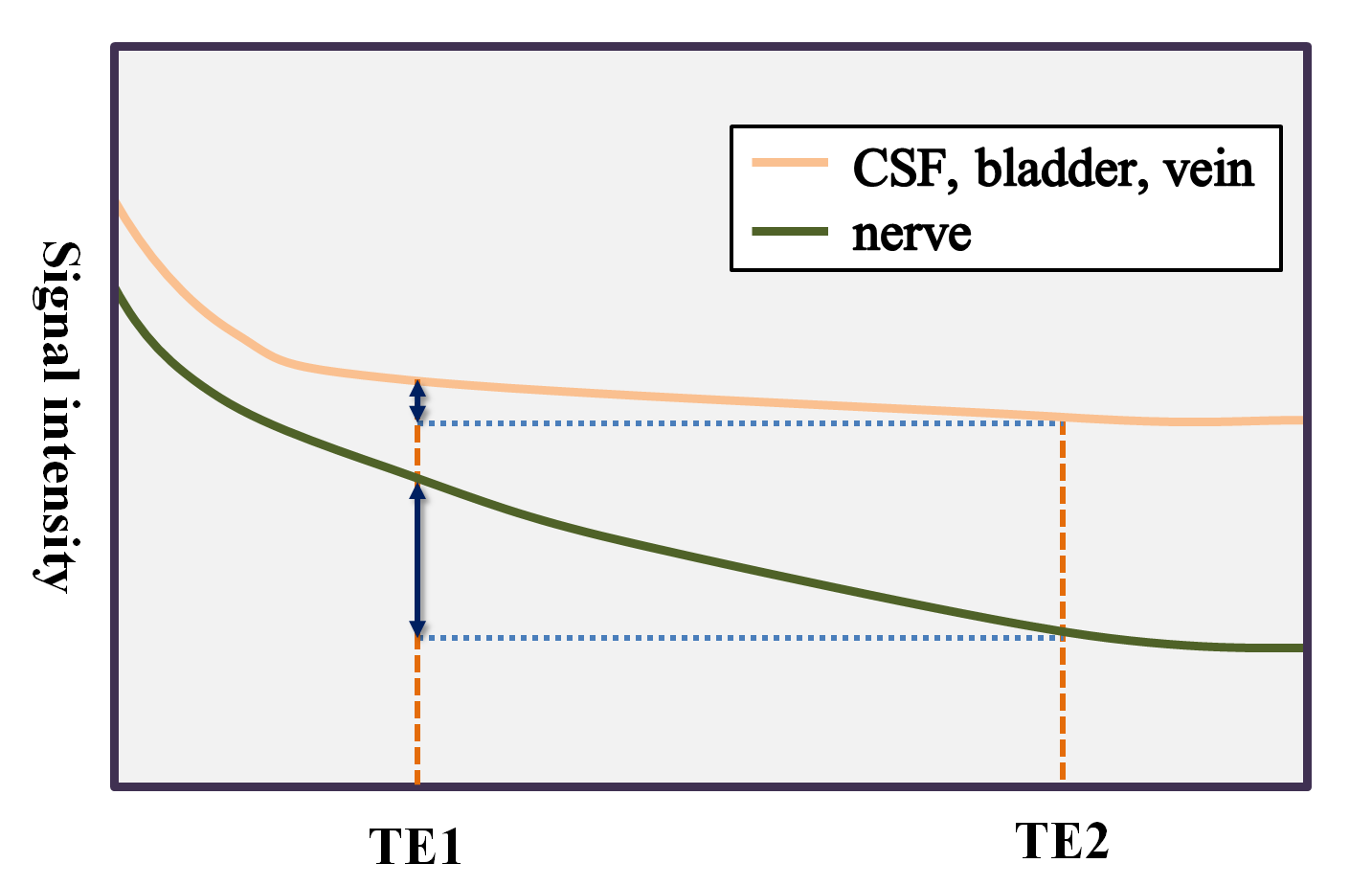
Fig. 1. HIRE requires two images with different TE to subtract signal
intensities of CSF, bladder, and veins. Each TE should be set to apply their
signal attenuation. In addition, the shorter TE should be set to demonstrate
nerves, and the longer TE should be set to show signal suppression of nerves.
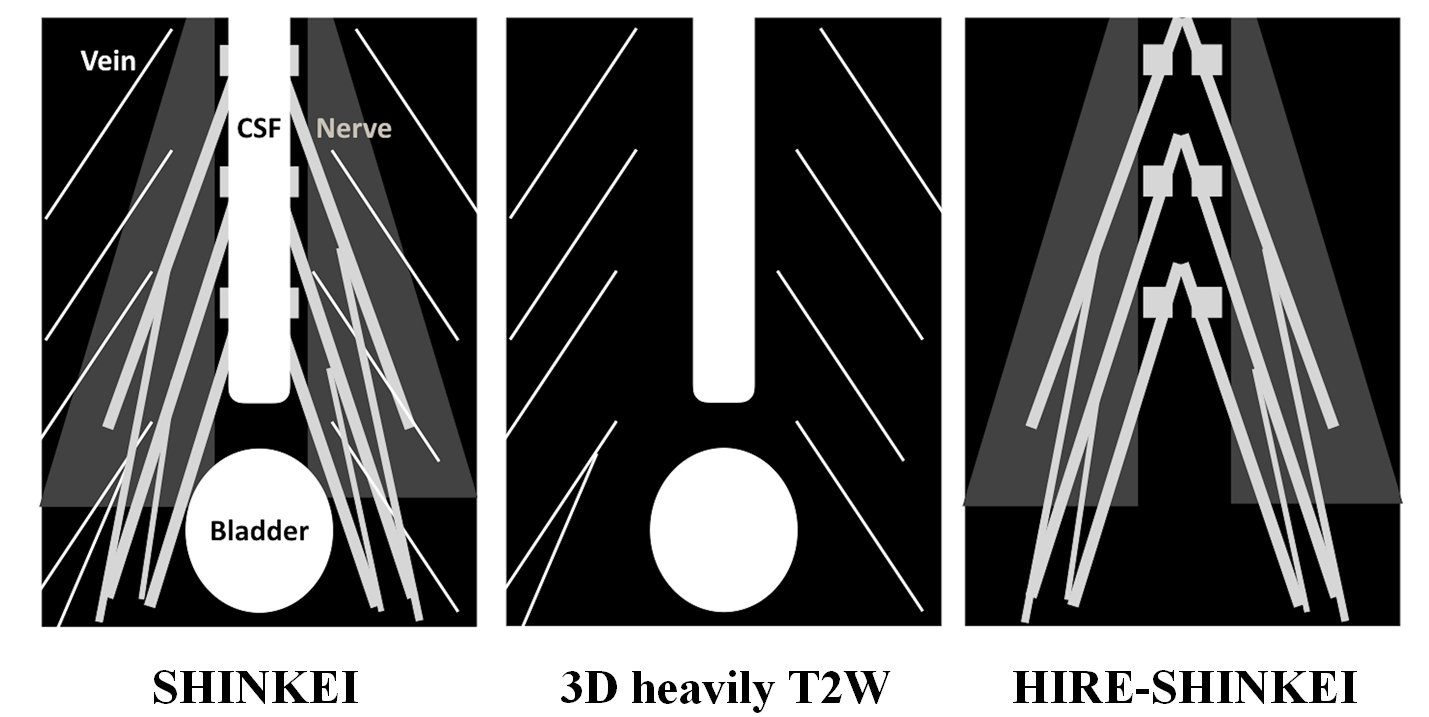
Fig. 2.
The scheme
of HIRE-SHINKEI. On HIRE-SHINKEI, 3D heavily T2W and SHINKEI are subtracted to
suppress signals of CSF, bladder, and veins. As a result, signals of nerves are
emphasized compared to SHINKEI.
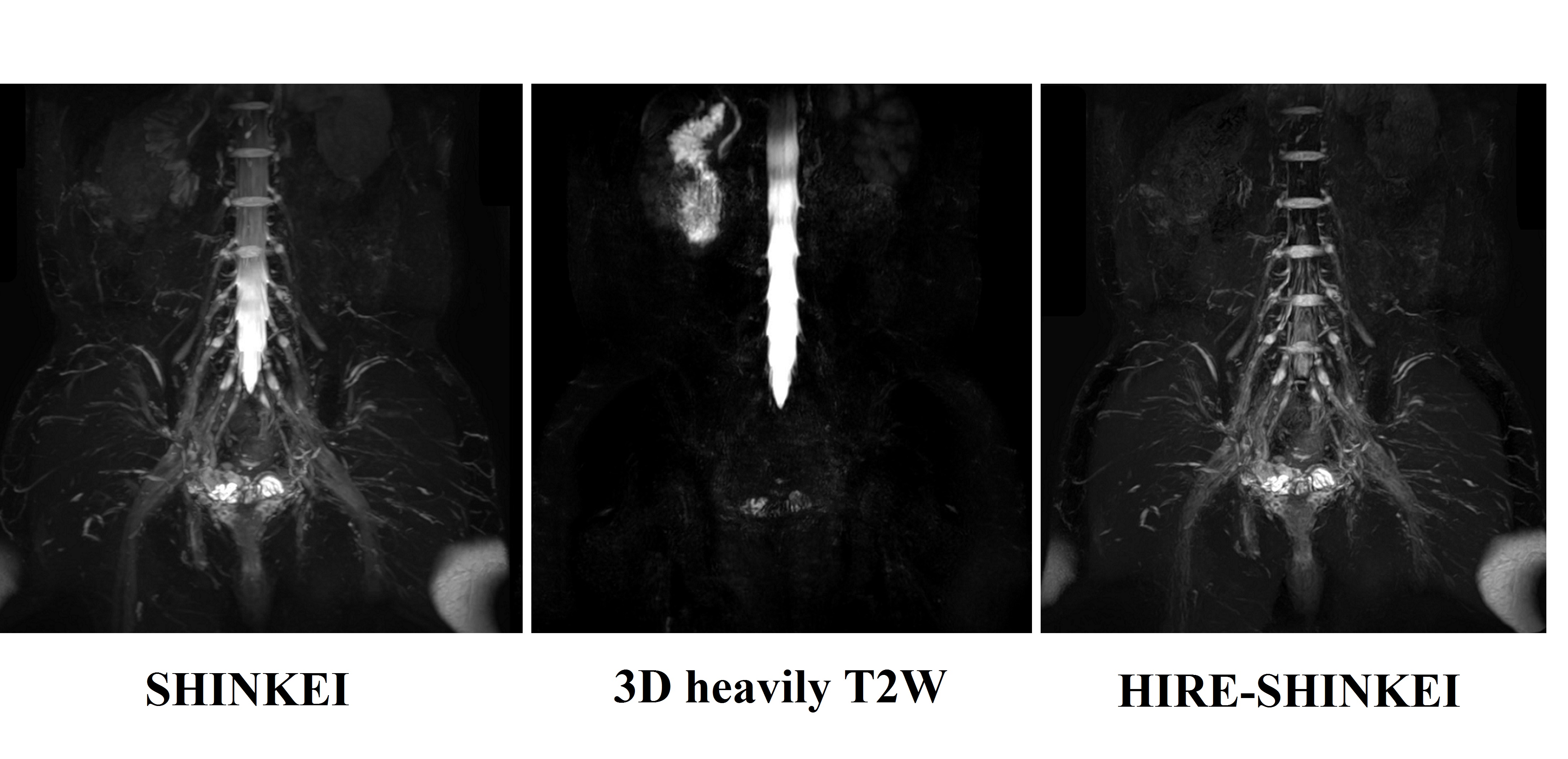
Fig.
3. MIP images of SHINKEI, 3D heavily T2W, and HIRE-SHINKEI.
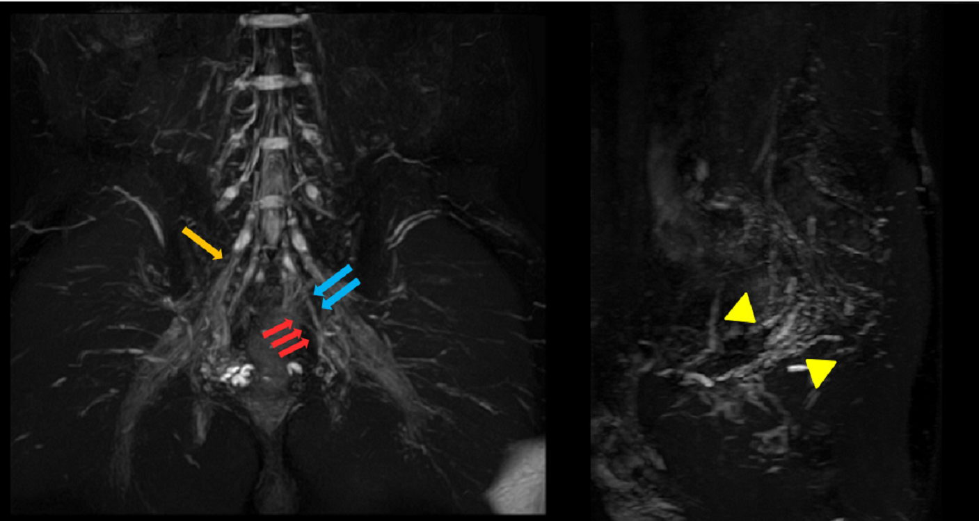
Fig. 4. MIP images on HIRE-SHINKEI. The origin of sacral
spinal nerves I (↓), II (↓↓), and III (↓↓↓) are clearly detected.
Pelvic splanchnic nerves (△) are clearly detected.