5138
3D variable flip angle fast spin echo imaging of the cervical spine : Improved image quality and decreased scan time with outer volume suppression1Radiology, Yonsei University College of Medicine, Seoul, Korea, Republic of, 2Global MR Applications & Workflow, GE Healthcare, Menlo Park, CA, 3GE Healthcare, Seoul, Korea, Republic of
Synopsis
Application of the volumetric fast spin echo (FSE) imaging in MRI is increasing, due to innate capability for multi-planar reconstruction, and potentials for a simplified workflow. However application to the cervical and thoracic area may be challenging because ghosting/phase encoded motion artifacts from swallowing, respiration and pulsations are frequent and severe in these regions, leading to image degradation. 3D FSE with outer volume suppression is a robust method for imaging the cervical spine, which provides improved image quality at a decreased scan time.
Purpose
Application of the volumetric fast spin echo (3D FSE) imaging in MRI is increasing, due to innate capability for multi-planar reconstruction, and potentials for a simplified workflow1.
Recently, various reduced FOV (field of view; rFOV)
methods have been introduced, and has been widely applied in spinal imaging,
especially for diffusion weighted images with EPI readouts, which considerably decreased distortion, leading to improved image quality.
Riffel et al. used inner volume excitation for lumbar spine imaging with phase-encoding(PE) in the AP direction, and addressed the potential for decreased scan time through decreased FOV and lack of need for oversampling, improved image quality by not using parallel imaging, and by elimination of artifacts from respiration and cardiac pulsation2.
However application to the cervical and thoracic area may be further challenging compared to the lumbar area, because ghosting/phase encoded motion artifacts from swallowing, respiration and pulsations are more frequent and severe in these regions, leading to image degradation. Remedies for conventional imaging include swap of frequency and phase encoding direction, or application of saturation pulses in the moving structures3.
We hypothesized that rFOV imaging with outer volume suppression(OVS) for 3D FSE can improve image quality and decrease scan time, as compared to the conventional 3D FSE protocol.
Method
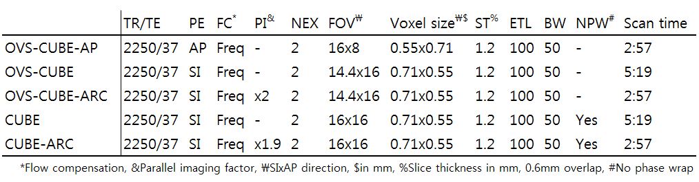
Two healthy volunteers were scanned on a 3T MR scanner (Discovery 750w, GE, Waukesha, WI) using a spine/neck array coil with 24 elements (GEM Spine, C Spine + Neck 24, GE, Waukesha, WI) in the sagittal plane, with the CUBE sequence, the GE variant of 3D variable flip angle FSE, with and without outer volume suppression. The scans were performed in random order and the detailed parameters are shown in Table 1.. The phase encoding directions were employed in the SI direction, except for OVS-CUBE-AP. Saturation bands were positioned covering the mouth, and pharyngo-esophageal areas. Flow compensation was only possible in the frequency direction due to the restraints within the sequence. For the OVS, the number of very selective saturation (VSS) pulses was 3.
Further technical details regarding the implemented OVS method, are described in detail elsewhere4.
For preliminary evaluation, the images were visually assessed by one radiologist with 5 years of experience in spine MRI.
Result
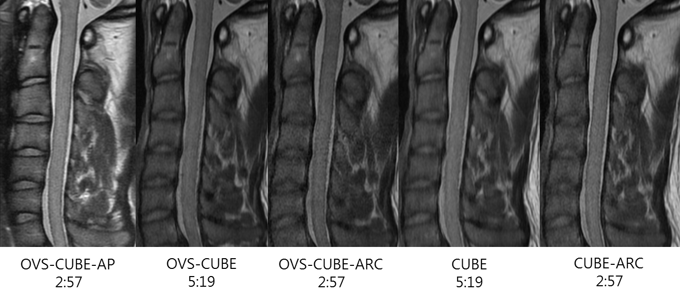
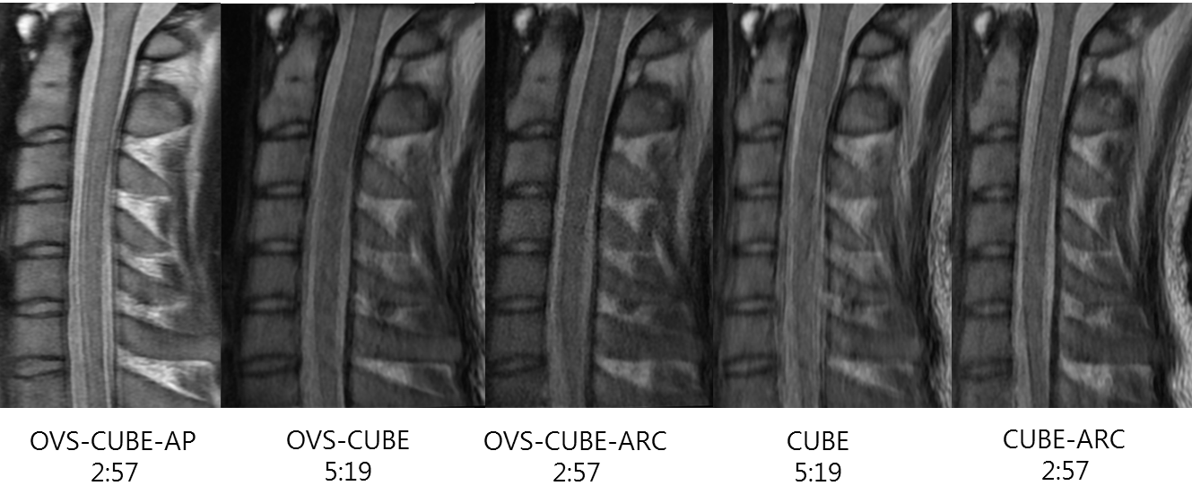
The image quality of the OVS-CUBE with phase encoding in the AP direction, showed superior image quality in terms of noise, image blurring and artifact as compared to the conventional imaging as well as OVS-CUBE with phase encoding in the SI direction (Figure 1, 2.).
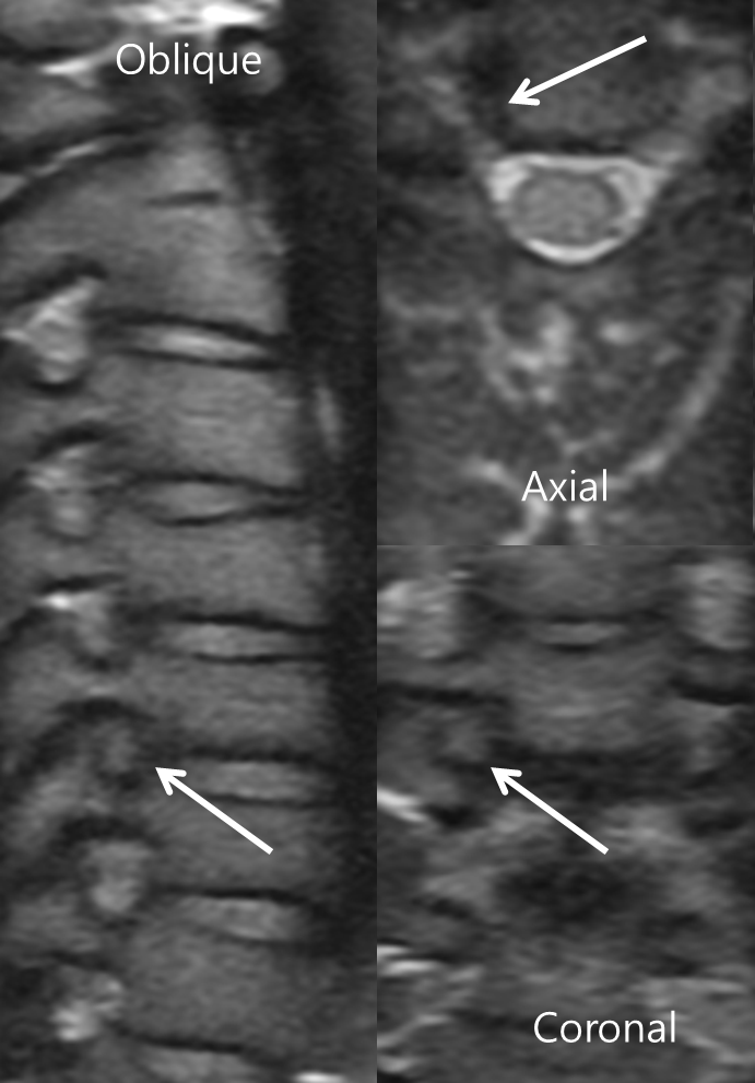
The quality of the oblique sagittal, axial and coronal reformatted images were also acceptable (Figure 3.).
Discussion
With the help of the reduced FOV capability enabled high resolution 3D FSE based acquisitions in the Cspine in less than 3 minutes, from which reformats in other planes could also be obtained with diagnostic quality. But since volumetric scans are sensitive to motion anytime during the scan, techniques to further accelerate the acquisition and better motion compensation techniques will be investigated.Conclusion
3D FSE with OVS is a robust method for imaging the cervical spine, which provides improved image quality at a decreased scan time.Acknowledgements
The authors thank Minsoo Park for his assistance in the protocol setup.
References
1. Sayah A, Jay AK, Toaff JS, Makariou EV, Berkowitz F. Effectiveness of a Rapid Lumbar Spine MRI Protocol Using 3D T2-Weighted SPACE Imaging Versus a Standard Protocol for Evaluation of Degenerative Changes of the Lumbar Spine. Am J Roentgenol. 2016;207(3):614-620.
2. Riffel P, Michaely HJ, Morelli JN, et al. Fast inner-volume imaging of the lumbar spine with a spatially focused excitation using a 3D-TSE sequence. Acad Radiol. 2015;22(4):423-429.
3. Morelli JN, Runge VM, Ai F, et al. An image-based approach to understanding the physics of MR artifacts. Radiographics. 2011;31(3):849-866
4. S Banerjee, M Han, W Chen, C Hess, R Krug, A Shankaranarayanan, Y Zur, Reduced Field-Of-View Imaging with 3D Variable Flip Angle Fast Spin Echo-Feasibility in MRI of Orbits. ISMRM 23rd Annual Meeting, Toronto, Canada, 2015, 2309
Figures