5136
Whole body FDG PET/MRI for assessment of facet joint osteoarthritis: Direct comparison of FDG uptake with MRI and CT features1Department of Radiology, Hyogo Cancer Center, Akashi, Japan, 2Department of Radiology, Kobe University Graduate School of Medicine, Kobe, Japan, 3Department of Radiology, National Cerebral and Cardiovascular Center, Suita, Japan
Synopsis
Hybrid PET/MRI scanner is an emerging modality and utilized not only for oncological or neurological purposes but also for assessment of musculoskeletal disease. Simultaneous acquisition of PET and MRI enables precise anatomical localization of PET tracers and further understanding of mechanism of the tracer uptake by using MRI information. Our hypothesis was that PET/MRI can reveal the pathophysiological mechanism of facet joint osteoarthritis by the simultaneous whole body imaging. The results showed MRI features on PET/MR significantly correlated with FDG uptake in the facet joint osteoarthritis and may be superior to CT based evaluation.
PURPOSE
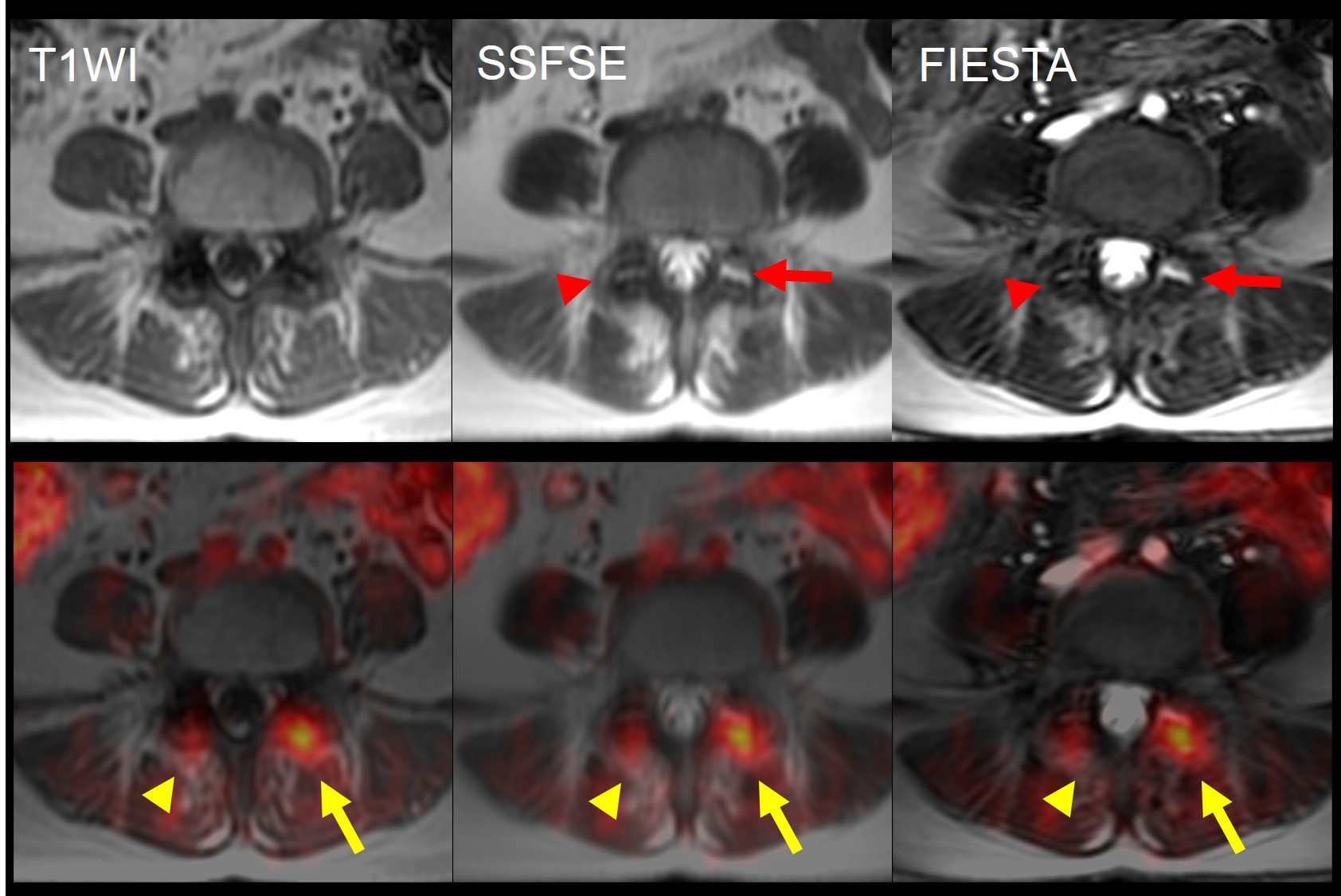
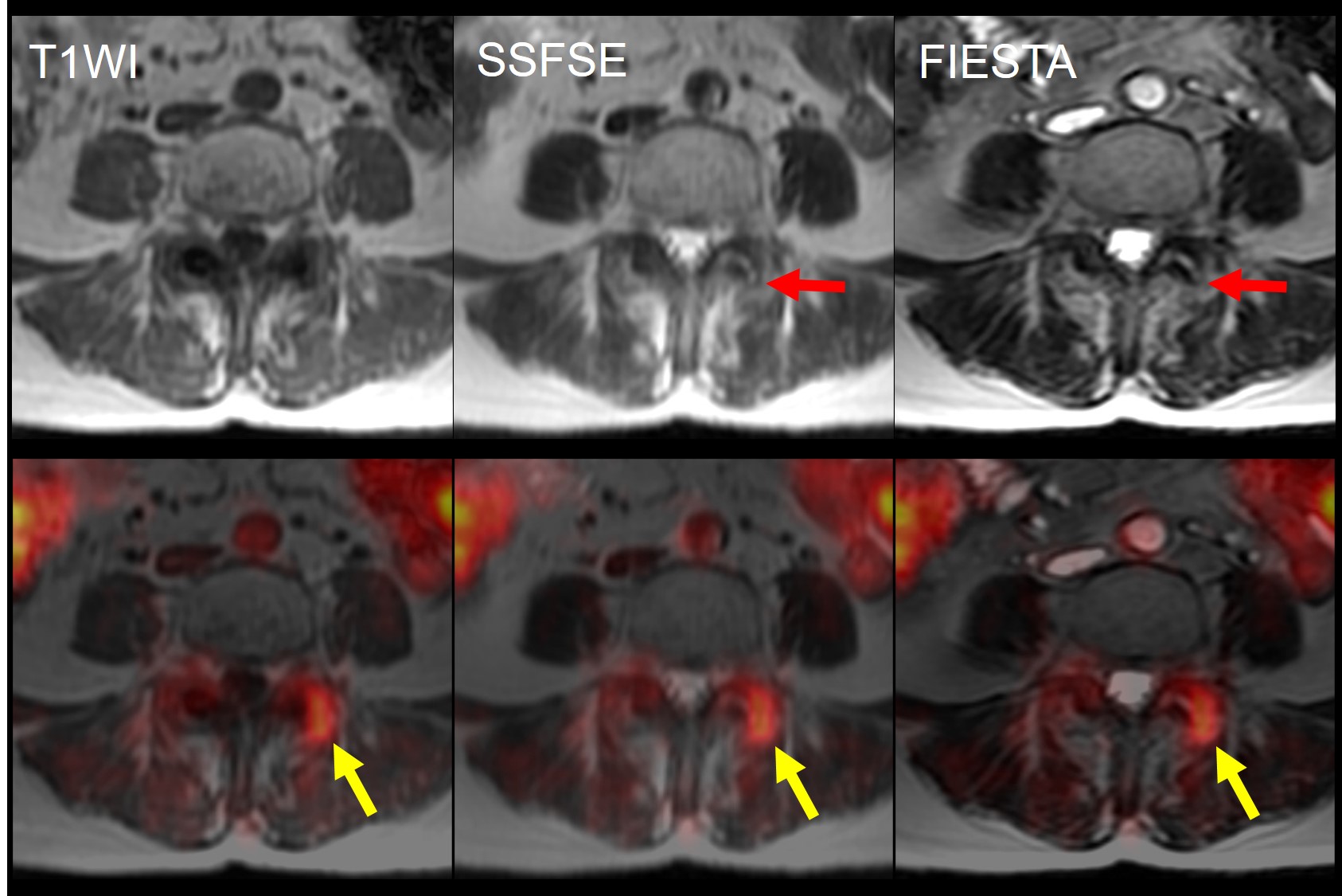
To evaluate the 2-[18F]Fluoro-2-deoxy-d-glucose (FDG) uptake of facet joints of the spine on whole body positron emission tomography/ magnetic resonance imaging (PET/MRI) and assess the correlation between tracer uptake and degenerative change on MRI and CT.MATERIALS AND METHODS
One hundred and twenty-two consecutive patients with clinically confirmed spondylosis deformans who underwent FDG PET/MRI for assessment of oncological disorders were retrospectively evaluated. Twenty-four of 122 subjects met the inclusion criteria and enrolled into the study population: CT acquisition including whole spine within three months; no apparent or confirmed malignancies in the spine; no hematological disease. FDG was administrated 60 minutes before PET emission scan, and whole body MRI including an axial whole body T1-weighted 3-dimensional dual-echo fast spoiled gradient-echo sequence (LAVA flex), an axial T2-weighted half-fourier acquisition single shot fast spin-echo (SSFSE) and an axial balanced steady-state gradient echo sequence (FIESTA) were simultaneously acquired on a 3.0 T hybrid PET/MRI scanner. Maximum standardized uptake value (SUVmax) were measured for all the facet joints on each subject by placing a region of interest on PET/MRI fusion images, and the anatomical location of the highest uptake voxel was recorded. MRI and CT features of the facet joints were visually evaluated by a board-certified radiologist using 3-point scoring system as follows: sclerotic change; hypertrophy of the joint and fluid collection within joint space. To evaluate the correlation between glucose metabolism on PET and anatomical features on MRI and CT, SUVmax and visual scores were statistically compared by the Spearman’s rank correlation and the multiple regression test. To assess the relationship between increased glucose metabolism and anatomical location in the joint, the SUVmax and the location of highest voxel were statistically compared by the one-way ANOVA.RESULTS
MRI and CT findings of hypertrophy and MRI finding of fluid collection showed significant positive correlation with SUVmax (p=0.0004 and 0.0026, respectively); however, no correlation was found between MRI and CT findings of sclerotic changes and SUVmax. On the multiple regression analysis, MRI finding of fluid collection was the only factor that significant correlated with SUVmax (p<0.0001). The highest FDG uptake was predominantly found in hypertrophic changes of superior and inferior articular process rather than joint space (p<0.05).CONCLUSIONS:
In this preliminary study, there is significant correlation between FDG uptake and MRI findings especially for fluid collection in the joint, suggesting the utility of MRI feature to clarify the glucose metabolism of facet joint osteoarthritis. Simultaneously acquired PET/MRI enables precise localization of FDG uptake in the facet joint, yielding better characterization of osteoarthritis.Acknowledgements
No acknowledgement found.References
1. Farshad-Amacker NA, Farshad M, Winklehner A, Andreisek G. MR imaging of degenerative disc disease. European journal of radiology 2015;84(9):1768-1776.
2. Gellhorn AC, Katz JN, Suri P. Osteoarthritis of the spine: the facet joints. Nature reviews Rheumatology 2013;9(4):216-224.
3. Kogan F, Fan AP, McWalter EJ, Oei EH, Quon A, Gold GE. PET/MRI of metabolic activity in osteoarthritis: A feasibility study. Journal of magnetic resonance imaging : JMRI 2016.
4. Weishaupt D, Zanetti M, Boos N, Hodler J. MR imaging and CT in osteoarthritis of the lumbar facet joints. Skeletal radiology 1999;28(4):215-219.
5. White ML, Johnson GB, Howe BM, Peller PJ, Broski SM. Spectrum of Benign Articular and Periarticular Findings at FDG PET/CT. Radiographics : a review publication of the Radiological Society of North America, Inc 2016;36(3):824-839.
Figures