5133
Correlation Between Bone Marrow Fatty Deposition Under Vertebral Endplates with Lumbar Intervertebral Disc Degeneration1Department of medical radiology, The Third Affiliated Hospital of Southern Medical University(Academy of orthopedics •Guangdong Province), Guangzhou, People's Republic of China, 2Philips Healthcare, Guangzhou, People's Republic of China
Synopsis
The intervertebral disc without blood supply obtained nutrient substance diffusing from the microcirculation under vertebral endplates. Some studies showed the fat deposition of bone marrow may pressure vessels and blood sinus, which could affect the microcirculation. So the fatty content of bone marrow under vertebral endplates can reflect the degeneration of the vertebral. However, the correlation of fatty content of bone marrow with lumbar intervertebral disc degeneration is not clear. The purpose of our study was to analyze the correlation between bone marrow fatty fraction under vertebral endplates with Pfirrmann Grades and T2* value of lumbar intervertebral disc.
Introduction
The intervertebral disc without blood supply obtained nutrient substance diffusing from the microcirculation under vertebral endplates. Some studies showed the fat deposition of bone marrow may pressure vessels and blood sinus, which could affect the microcirculation. So the fatty content of bone marrow under vertebral endplates can reflect the degeneration of the vertebral. However, the correlation of fatty content of bone marrow with lumbar intervertebral disc degeneration is not clear. The purpose of our study was to analyze the correlation between bone marrow fatty fraction under vertebral endplates with Pfirrmann Grades and T2* value of lumbar intervertebral disc.Methods
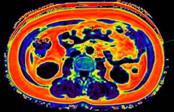
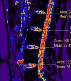
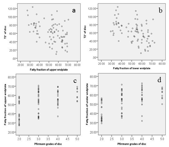
This study was approved by our local ethics committee. 20 patients (with mean age of 54 years) with low back pain were recruited into the study. All patients underwent conventional spinal MRI concluding sagittal T2WI, axial mDixon-Quant protocol and sagittal T2* Mapping on a 3.0T scanner (Ingenia 3.0T, Philips Healthcare, Best, The Netherlands) with a 44-channel dStream spinal coil. The mDixon-Quant protocol was performed with FFE T1WI (six echo, TR: shortest, TE: shortest, FOV: 400*350, matrix: 176*174), and the T2* Mapping was performed with FFE T2*WI (16 echo, TR: shortest, TE: shortest, FOV: 350*350, matrix: 176*174). All data were transferred to the post processing workstation. The fatty fraction (FF) of bone marrow under upper and lower vertebral endplates and T2* values of L1/2-L4/5 intervertebral disc were measured. The degeneration of the discs were classified according to the Pfirrmann grading on sagittal T2-weithted images. The FF value of bone marrow and T2* value of disc between the different Pfirrmann grades were compared statistically by using one-way analysis of variance (ANOVA). The Pearson correlation of the FF value of bone marrow with the T2* value of disc and the Spearman correlation of the FF value of bone marrow with Pfirrmann grades of the lumbar intervertebral disc were analyzed. P value <0.05 was considered significant.Results and Discussion
The FF value of bone marrow and T2* value of disc between the different Pfirrmann grades showed significant difference (P <0.05). The FF value of bone marrow under upper or lower vertebral endplates were both significantly negative correlated with T2* value of the disc ( the correlation coefficient were -0.467 and-0.483,p<0.001). In addition, the FF value of bone marrow under upper or lower vertebral endplates was also significantly positive correlated with the Pfirrmann grades of disc degeneration(the correlation coefficient were 0.517 and 0.574,p<0.001.Conclusion
The fatty fraction of bone marrow under vertebral endplates measured by mDixon-Quant method correlates with the degree of lumbar intervertebral disc degeneration. The more fat deposition of bone marrow under vertebral endplates were,the more serious degeneration of the lumber intervertebral disc occurred.Acknowledgements
No acknowledgement found.References
[1] Castro-Mateos I, et al. Eur Spine J. 2016 Sep;25(9):2721-7.
[2] Yoon MA, et al. Magn Reson Imaging. 2016 Sep;34(7):932-9.
[3] Shen S, et al. Iran J Radiol. 2016 Jan 16;13(1):e30069. [4] Urrutia J, et al. Eur Spine J. 2016 Sep;25(9):2728-33.
[5] Bolan PJ, et al. J Magn Reson Imaging. 2013 Dec; 38(6):1578-84.
Figures