5125
Age-related changes in healthy thigh musculature: Multi-parametric MR imaging analysis1Department of Radiology, Korea University Guro Hospital, Seoul, Korea, Republic of, 2Department of Radiology, Korea University Anam Hospital, Seoul, Korea, Republic of, 3Korea University Ansan Hospital, Gyeonggi-do, Korea, Republic of, 4Siemens Healthcare Korea, Seoul, Korea, Republic of
Synopsis
Purpose: To identify MR parameters that are significantly associated with aging in healthy thigh muscles and investigate age-related characteristics.
Methods: Thirty healthy subjects underwent multi-parametric MRI of the right thigh, including IVIM-DWI, DTI, multi-echo Dixon imaging, DCE-MRI. Parametric maps of the thigh muscles (divided into three compartments) were analyzed. Relationships between parameters and age were assessed with simple and multiple linear regression analyses.
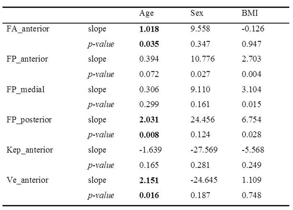
Results: Fractional anisotropy (slope=1.018) and Ve (slope=2.151) in anterior compartment and fat percentage in posterior compartment (slope=2.031) showed significant relationships to age (p<0.05).
Conclusion: Several MRI parameters of the thigh muscles were associated with normal aging.
Purpose
MRI evaluation of skeletal muscle is often challenged by normal aging-related changes. As a consequence of normal aging, skeletal muscles show structural alterations, including increased intramuscular fatty infiltration, fibrosis, decrease in type II muscle fiber size and number, fiber type grouping and atrophy.1-5 Moreover, skeletal muscles show diminished perfusion and declined oxidative capacity with increased aging.3,4 Thus, considerations for changes in skeletal muscle with normal age-related processes are necessary in evaluation of pathophysiology of muscle disease. Recently, several researches have shown the potential of quantitative MRI techniques as imaging biomarkers in evaluation of skeletal muscle diseases.6-8 They include intravoxel incoherent motion diffusion weighted imaging (IVIM-DWI), diffusion tensor imaging (DTI), multi-echo Dixon imaging and dynamic contrast enhanced (DCE)-MRI. IVIM-DWI provides information on both true water diffusion and tissue perfusion without need for contrast agent. DTI is sensitive to the directionality of water motion, thus can reflect muscle microstructural alterations.7 Multi-echo Dixon has shown comparable results to MR spectroscopy in quantification of skeletal muscle fat,8 and has advantages over MR spectroscopy in tissues with uneven fat distribution like skeletal muscles. Finally, DCE-MRI allows quantifications of changes in vessel permeability, blood flow and extracellular space.9 Each quantitative MRI technique mentioned above targets different components of the skeletal muscle and may provide complementary information on muscle pathophysiology. Therefore, the primary aim of the present study is to investigate age-related characteristics of thigh muscles in healthy subjects by applying multi-parametric quantitative MRI and identify MR parameters that are significantly associated with aging.Methods
Subjects: Thirty consecutive healthy subjects (15 men, 15 women; mean age ± SD: 46.87 ± 10.51 years; range 30-62 years) with normal or healthy weight (mean BMI ± SD: 22.96 ± 2.27) were included in this prospective study. No subject had muscle disorder, diabetes, cardiovascular, neurological disorders, histories of operation or trauma in the thigh region or was on medication. Age groups as follows: 30-39 years (n=9), 40-49 years (n=5), 50-59 years (n=14), 60-69 years (n=2).
Image acquisition: All patients underwent multi-parametric MRI of the right thigh on 3.0T scanner (Magnetom Skyra; Siemens, Erlangen, Germany). IVIM-DWI was acquired using single-shot echo planar imaging (SSEPI) with SPAIR with b-values (b=0, 25, 50, 75, 100, 200, 500, 800 s/mm2), TR/TE=340/60 ms, voxel size=1.3x1.3x5.0 mm3. For DTI, SSEPI with SPAIR was used with voxel size=2.5x2.5x5.0 mm3, TR/TE = 3300/60.0 ms, b-values=0, 450 s/mm2, 20 diffusion weighted directions. Multi-echo three-dimensional Dixon CAIPIRINHA-VIBE with T2*-correction was performed with TR = 9.15 ms, TE = 1.09, 2.46, 3.69, 4.92, 6.15, 7.38 ms, FA = 4°, voxel size 1.1x1.1x5.0 mm3. DCE-MRI was acquire using CAIPIRINHA-TWIST-VIBE with a temporal resolution of 4.94s, TR/TE=4.90/2.46 ms, FA=9°, voxel size=0.8x0.8x5.0 mm3, after 0.2mL/kg body weight gadolinium Gd-Dota (Dotarem, Guerbet, Aulnay-Sous-Bois, France), followed by 25mL saline flush. After pre-contrast T1 mapping (FA=2, 8, 15°), DCE-MRI was acquired with acquisition time of 10.03 m (120 phases). Population averaged AIFs with the lowest Chi2 was used for pharmacokinetic perfusion analyses. For all multi-parametric imaging, the same FOV of 220 mm, slice thickness of 5 mm with 24 slices were used.
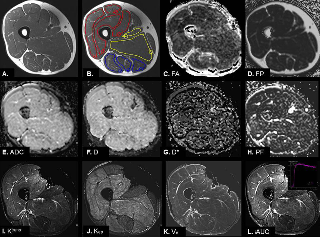
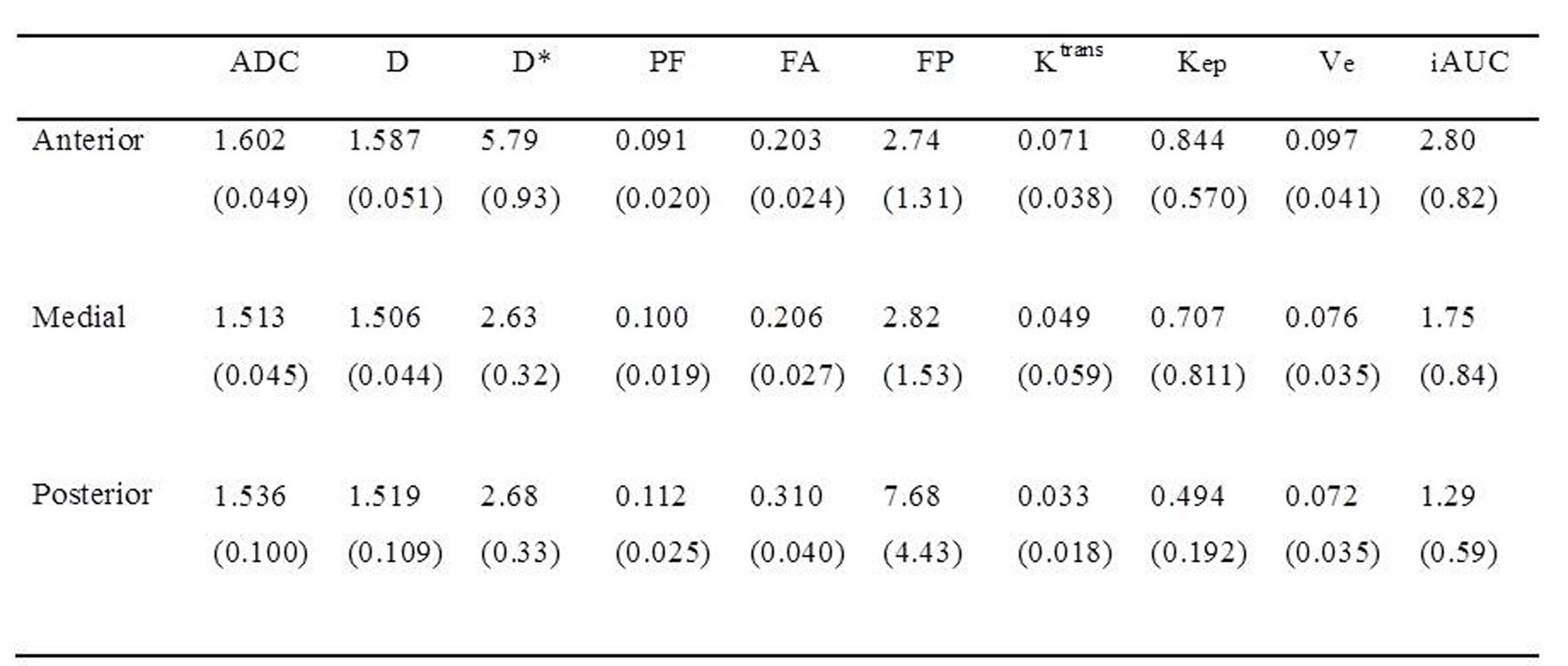
Image analyses: Measurements of parametric maps were made on the three consecutive axial images at the mid femur shaft level. ROIs were drawn to encompass each muscle, while avoiding gross visible blood vessels in the following thigh muscles: (1) anterior compartment: rectus femoris, vastus lateralis, medialis and intermedius; (2) medial compartment: adductor longus and magnus; (3) posterior compartment: biceps femoris, semitendinosus and semimembranosus. Mean values from three measurements were used for statistical analysis.
Statistical analyses: Relationships between parameters and age were assessed using simple linear regression analyses. Then, multiple linear regression analysis was performed to adjust for confounding from sex and BMI.
Results
Simple linear regression showed that fractional anisotropy (FA) in anterior compartment was positively correlated with age (r=0.553, p=0.002). Fat percentages in all three compartments showed positive correlation with age (anterior, r=0.570, p=0.001; medial, r=0.403, p=.027; posterior, r=0.655, p=0.000). Regarding DCE-MRI, Kep and Ve in anterior compartment were significantly associated with age (r=-448, p=0.013 and r=0.390, p=0.033, respectively). IVIM-DWI did not show significant relationships to age. When adjusted for confounding from sex and BMI by multiple linear regression analyses, FA in anterior compartment, fat percentage in posterior compartment and Ve in anterior compartment showed significant relationships to age.Conclusion
Our study suggests that a number of MRI parameters of the thigh muscles (FA, fat percentage and Ve) are associated with normal aging. Changes in these parameters may reflect microstructural, histologic and microcirculatory changes within skeletal muscle with advancing aging.Acknowledgements
No acknowledgement found.References
1. Andersen JL. Muscle fibre type adaptation in the elderly human muscle. Scand J Med Sci Sports. 2003;13:40-47.
2. Csapo R, Malis V, Sinha U, et al. Age-associated differences in triceps surae muscle composition and strength - an MRI-based cross-sectional comparison of contractile, adipose and connective tissue. BMC Musculoskelet Disord. 2014;15:209.
3. Wray DW, Nishiyama SK, Monnet A, et al. Antioxidants and aging: NMR-based evidence of improved skeletal muscle perfusion and energetics. Am J Physiol Heart Circ Physiol. 2009;297:H1870-1875.
4. Haseler LJ, Lin AP, Richardson RS. Skeletal muscle oxidative metabolism in sedentary humans: 31P-MRS assessment of O2 supply and demand limitations. J Appl Physiol (1985). 2004;97:1077-1081.
5. Korhonen MT, Cristea A, Alen M, et al. Aging, muscle fiber type, and contractile function in sprint-trained athletes. J Appl Physiol (1985). 2006; 01:906-917.
6. Filli L, Boss A, Wurnig MC, et al. Dynamic intravoxel incoherent motion imaging of skeletal muscle at rest and after exercise. NMR Biomed. 2015;28:240-246.
7. Oudeman J, Nederveen AJ, Strijkers GJ, et al. Techniques and applications of skeletal muscle diffusion tensor imaging: A review. J Magn Reson Imaging. 2016;43:773-788.
8. Agten CA, Rosskopf AB, Gerber C, et al. Quantification of early fatty infiltration of the rotator cuff muscles: comparison of multi-echo Dixon with single-voxel MR spectroscopy. Eur Radiol. 2016;26:3719-3727.
9. Verma S, Turkbey B, Muradyan N, et al. Overview of dynamic contrast-enhanced MRI in prostate cancer diagnosis and management. AJR. 2012;198:1277-1288.
Figures