5120
Accuracy of Age Estimation based on Undersampled MR Images of the Hand1Ludwig Boltzmann Institute for Clinical Forensic Imaging, Graz, Austria, 2BioTechMed-Graz, Graz, Austria, 3Institute of Medical Engineering, Graz University of Technology, Graz, Austria, 4Institute for Computer Graphics and Vision, Graz University of Technology, Graz, Austria
Synopsis
The topic of age estimation in living persons is important for sports competitions as well as unaccompanied minors without valid documents and has recently gained interest. Recommended imaging protocols are based on modalities using ionizing radiation and therefore MRI is currently investigated for its eligibility to replace these measurements. This study investigates the accuracy of MRI-based age estimation for retrospectively undersampled data to determine the degree of inaccuracies introduced by an acceleration of the acquisition technique using commercially available undersampling strategies. For this purpose the data is analyzed by a radiologist and using an automatic method.
Purpose
The estimation of the chronological age of living persons is important in sporting events with certain age limits and especially for unaccompanied adolescents without valid documents as a person’s age has legal consequences when entering a country.
As current recommendations1 suggest using ionizing radiation-based imaging modalities, the interest in MRI has noticeably increased. Recently, an automatic estimation method2 was published and its precision was tested on undersampled data3. As short acquisition times are desirable for young subjects, first studies investigated the reliability of age estimates using data acquired with optimized undersampling patterns4.
This study investigates the accuracy of age estimation methods based on accelerated MR image acquisition with commercially available undersampling strategies by evaluation of different estimation and image reconstruction methods.
Methods
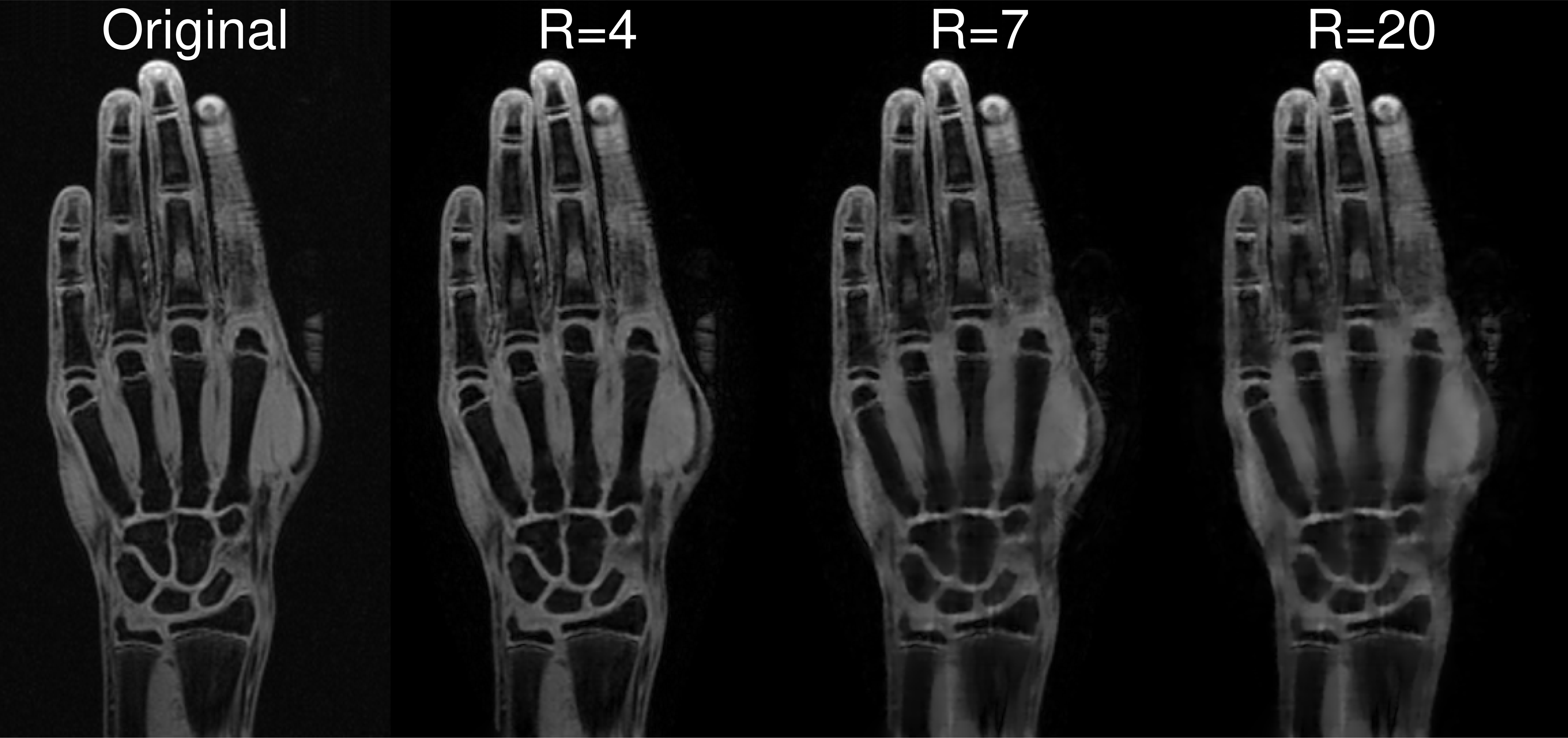
For ten healthy male volunteers between 13 and 19 years T1-weighted 3D FLASH VIBE measurements (parameters in Figure 1) of the left hand were acquired on clinical 3T MR scanners.
Non-averaged raw k-space data was undersampled according to the CAIPIRINHA strategy5 with 12 calibration lines and (rounded) acceleration factors $$$R\in\{4,7,11,14,17,20\}$$$. Image reconstruction was carried out with TGV6, and CG-SENSE7 using the AVIONIC8 and BART9 toolboxes, respectively.
A forensic radiologist with more than 5 years of experience was presented original images and images TGV-reconstructed from data undersampled with R=4 and R=7 (corresponding to acquisition times of 29 and 15 seconds, respectively) in a randomized manner and applied the Greulich-Pyle (G-P) method10 to estimate the subjects’ age. Automatic age estimation used the method proposed by Urschler et al.2 extended by improved landmark localization11 and a deep neural network based age estimator12. The automatic age estimation method was tested for all undersampling factors and image reconstruction methods.
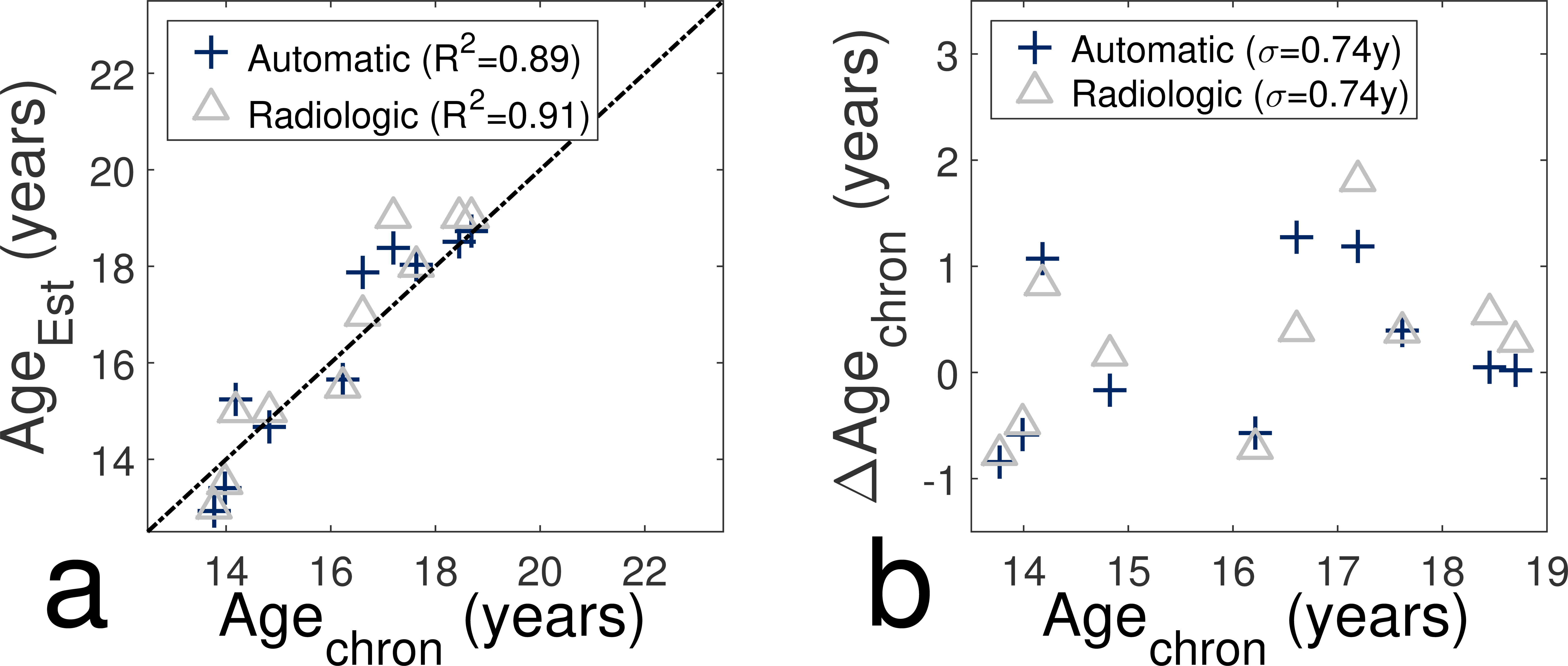
Accuracy of the results was evaluated by analyzing the correlation coefficient R2 of a linear regression between chronological and estimated age and the standard deviation σ of the difference between chronological and estimated age.
Results
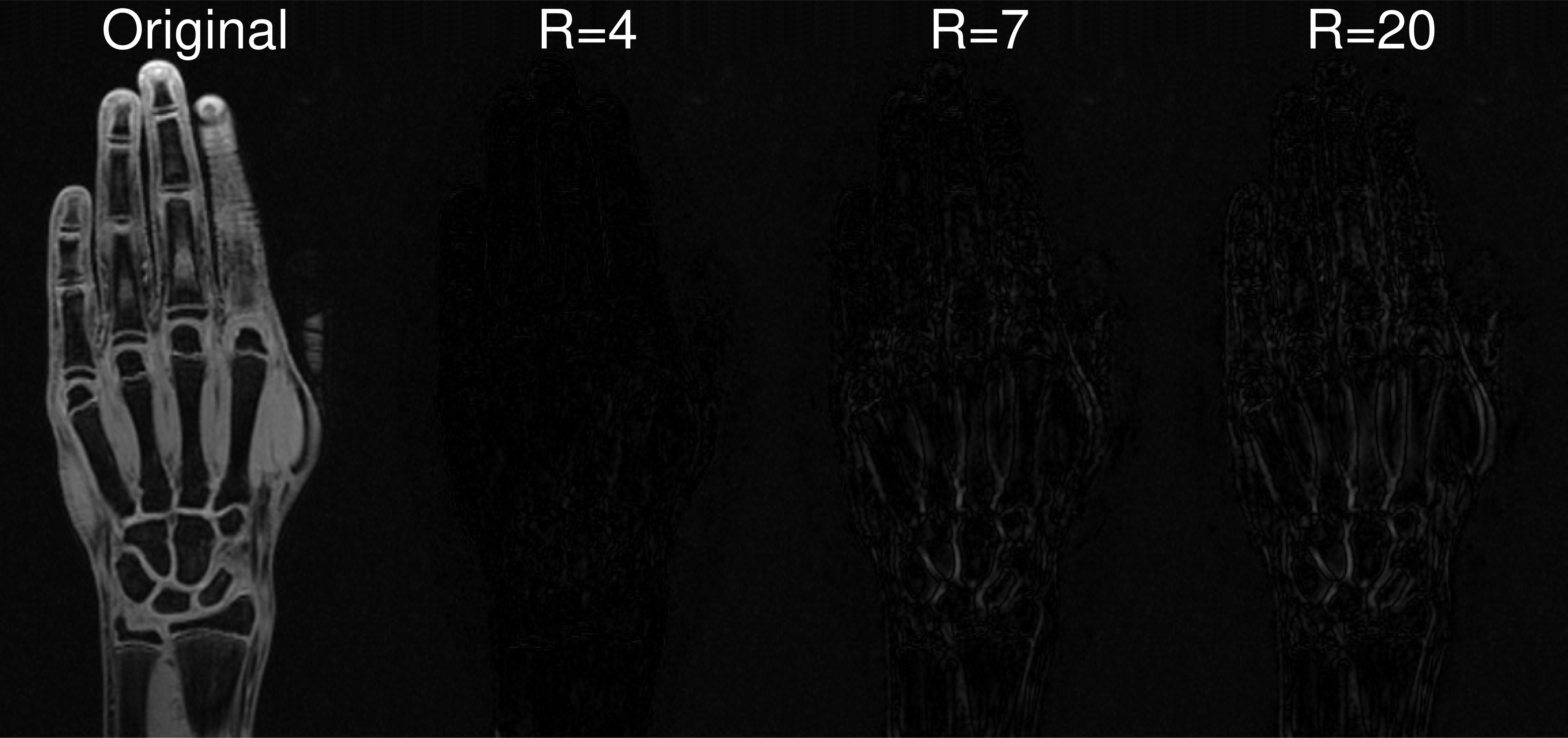
Figure 1 shows an exemplary slice of one volunteer for original data and TGV-reconstructions from data undersampled with R=4, 7 and 20, Figure 2 shows the original image and differences to the undersampled images in Figure 1.
Figure 3 shows the accuracy of the expert and the automatic age estimation method by comparing the results for the fully-sampled data sets; values for R2 and σ (always shown in figures) are almost identical.
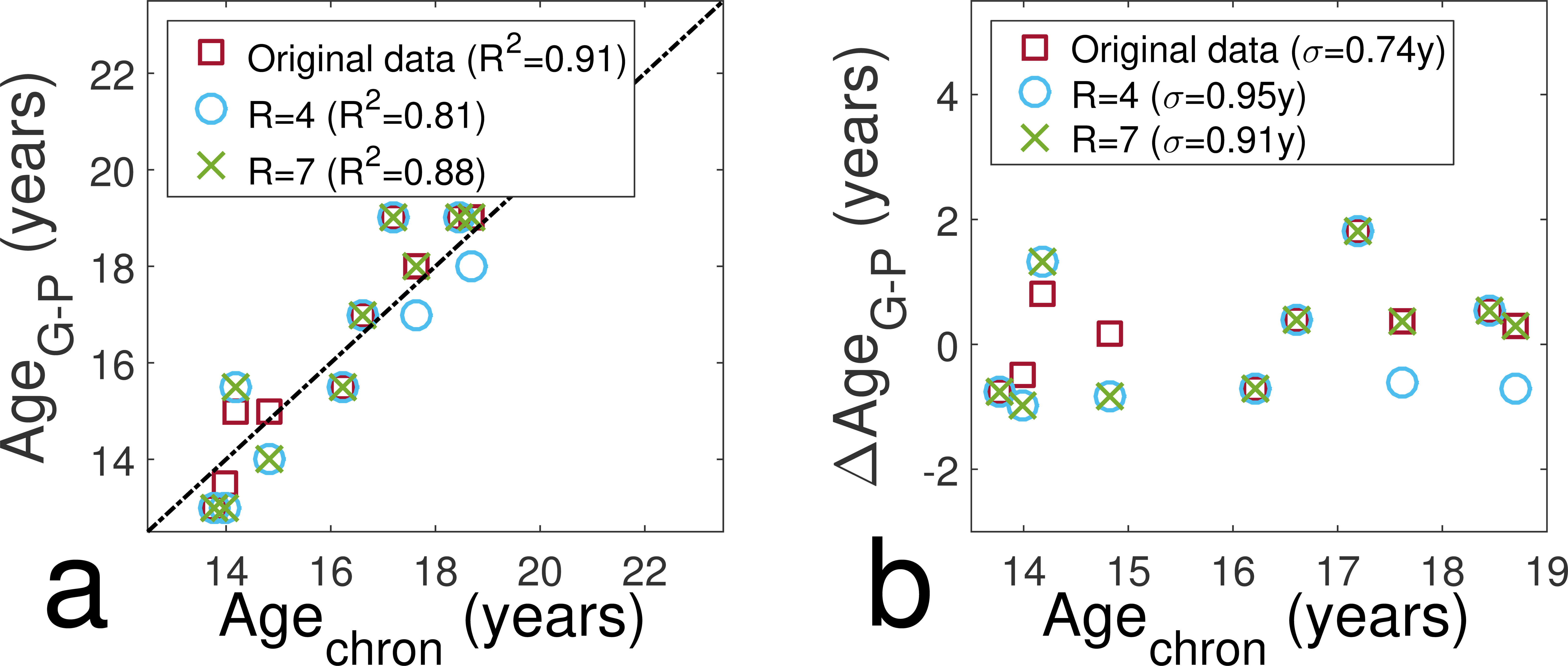
With increasing acceleration factor the agreement between chronological age and the age estimated by the radiologist decreases slightly (decrease of R2 and increase of σ), which is shown in Figure 4.
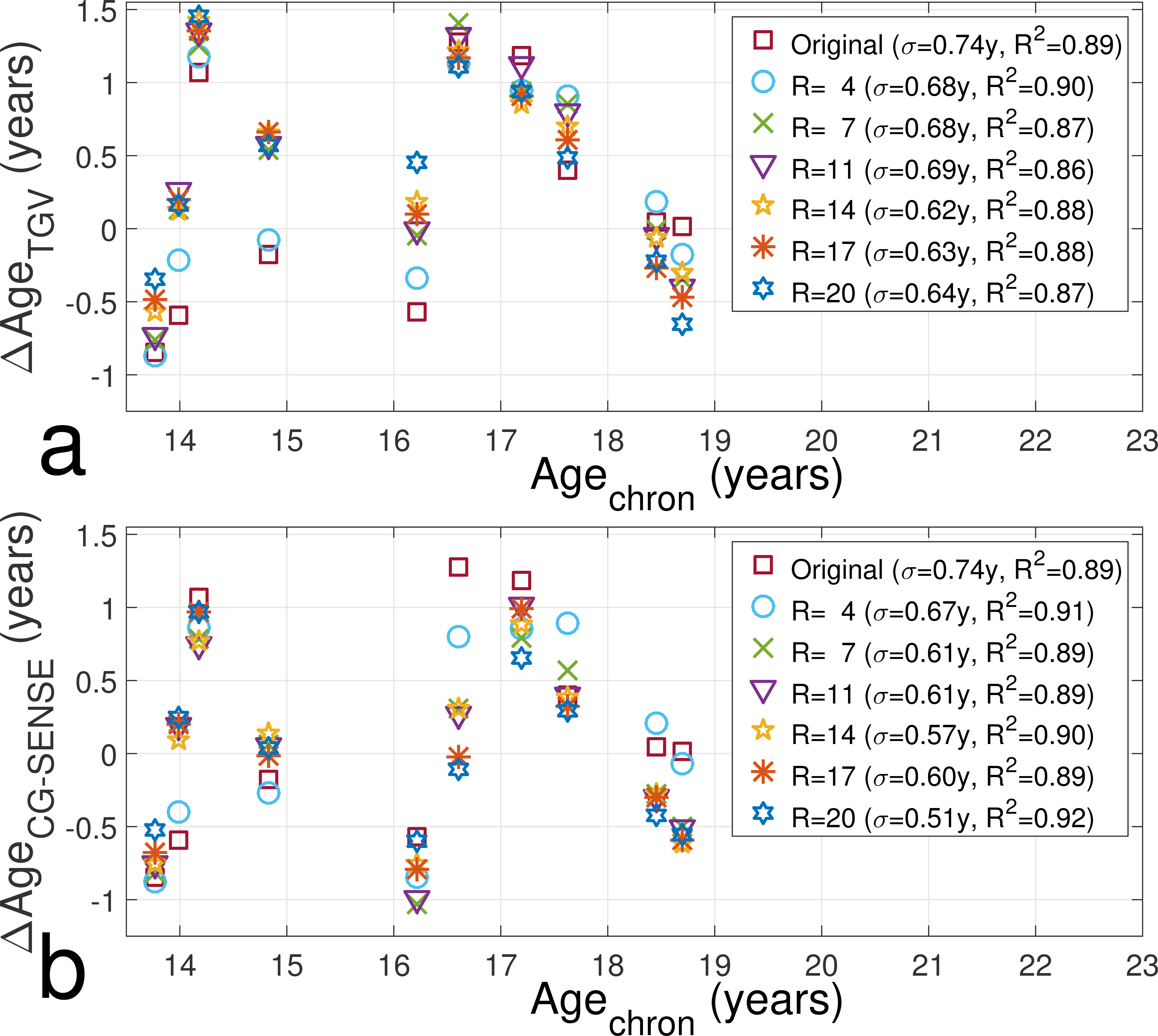
For the automatic age estimation (Figure 5) the accuracy of the age estimation did not decrease with increasing undersampling factor. The standard deviation of the differences did not exceed the value for the fully-sampled data set for both TGV and CG-SENSE reconstructions. The values for R2 were slightly decreased for TGV-reconstructions and equal to or higher than for fully-sampled data for CG-SENSE reconstructions.
Discussion and Conclusion
The aim of this study was to investigate the accuracy of age estimation methods based on MR images reconstructed from undersampled data to determine whether a speed-up of image acquisition introduces not acceptable errors into the age assessment process.
The difference between estimated and chronological age for the fully-sampled data sets is in the range of reported uncertainties for bone age estimation13 (0.5–2 years) and comparable for both the radiologic and automatic method, which forms an important basis for this study.
Images with R>7 were not presented to the radiologist as it was shown that these are mostly rated as not assessable14 and for all evaluated images the standard deviation was below 1 year. Due to the small number of volunteers only images reconstructed with one algorithm were presented to avoid biased age estimates. Higher subject numbers will also allow an acceptable randomization for more reconstruction techniques.
The remarkable result of this study is that even for high acceleration factors the undersampling has no determinable influence on the automatic age estimation. This can be explained by the fact, that the automatic algorithm analyzes the whole 3D data set. Thereby, the age estimation is not affected by single artefacts, which may e.g. mimic a physeal closure and influence the result of a radiologic analysis. Furthermore, the insensitivity to the reconstruction algorithm is also a sign for the robustness of the method.
Since age estimation is mainly performed for adolescents, short acquisition times are highly desirable. The preliminary results of this study will further promote the use of MRI for age estimation as they show that acquisition times can be remarkably reduced without a loss in accuracy.
Acknowledgements
This work was supported by the SFB "Mathematical Optimization and Applications in Biomedical Sciences'' of the Austrian Science Fund (FWF): SFB F3209-18 and the project FAME of the Austrian Science Fund (FWF): P28078-N33.References
1. A. Schmeling, C. Grundmann, A. Fuhrmann, et al. Updated recommendations of the Study Group on Forensic Age Diagnostics for age estimation in the living in criminal proceedings. In: Rechtsmedizin 18.6 (2008), pp. 451-453.
2. M. Urschler, S. Grassegger, and D. Stern. What automated age estimation of hand and wrist MRI data tells us about skeletal maturation in male adolescents. In: Ann Hum Biol 42.4 (2015), pp. 358–367.
3. B. Neumayer, M. Schloegl, C. Payer, et al. Acceleration of MR Measurements for Age Estimation. In: Magn Reson Mater Phy (2016) 29:325–326.
4. Y. Terada, D. Tamada, K. Kose, et al. Acceleration of skeletal age MR examination using compressed sensing. In: J Magn Reson Imaging 44.1 (2015), pp. 204–211.
5. F. A. Breuer, M. Blaimer, R. M. Heidemann, et al. Controlled aliasing in parallel imaging results in higher acceleration (CAIPIRINHA) for multi-slice imaging. In: Magn Reson Med 53.3 (2005), pp. 684–691.
6. F. Knoll, K. Bredies, T. Pock, and R. Stollberger. Second order total generalized variation (TGV) for MRI. In: Magn Reson Med 65.2 (2010),pp. 480–491
7. K. P. Pruessmann, M. Weiger, M. B. Scheidegger and P. Boesiger. SENSE: Sensitivity encoding for fast MRI. Magn. Reson. Med. (1999) 42: 952–962
8. M. Schloegl, M. Holler, A. Schwarzl, et al. Infimal convolution of total generalized variation functionals for dynamic MRI. Magn. Reson. Med. (2016) doi:10.1002/mrm.26352
9. M. Uecker, F. Ong, J. I. Tamir, et al. Berkeley Advanced Reconstruction Toolbox, In: Proc. Intl. Soc. Mag. Reson. Med. (2015) 23:2486
10. W. W. Greulich and S. I. Pyle. Radiographic atlas of skeletal development of the hand and wrist. In: Am J Med Sci 238.3 (1959), p. 393
11. D. Stern, T. Ebner, and M. Urschler. From local to global random regression forests: Exploring anatomical landmark localization. In: Med Image Comput Comput Assist Interv (2016).
12. D. Stern, C. Payer, V. Lepetit, and M. Urschler. Automated Age Estimation from Hand MRI Volumes using Deep Learning. In: Med Image Comput Comput Assist Interv (2016).
13. S. Ritz-Timme, C. Cattaneo, M. J. Collins, et al. Age estimation: The state of the art in relation to the specific demands of forensic practise. In: Int J Legal Med 113.3 (2000), pp. 129–136.
14. M. Schloegl, B. Neumayer, T. Ehammer, et al. Validation of TGV regularized accelerated MR reconstruction by age estimation. In: Magn Reson Mater Phy (2016) 29:104–105.Figures