5112
MRI study of the changes of perfusion and fat content in radiation-induced bone marrow injury in rats1Department Of Radiology, Renmin Hospital Of Wuhan University, wuhan, People's Republic of China, 2GE Healthcare, Shanghai, People's Republic of China
Synopsis
This study is to investigate whether adipocytes influence bone marrow microcirculation based on DCE-MRI and ex vivo high-resolution MAS 1H NMR spectroscopy. In this animal review committee-approved study, Rat Femurs were evaluated through DCE-MRI, 1H HRMAS NMRS, and histopathologic analysis before irradiation and on days 4 and 7 after irradiation. The results showed Ktrans and adipocytes gradually increased a peak on the 7th day after irradiation. In conclusion, bone marrow microcirculation permeability correlation with fat content after irradiation suggested for the first time that a pathophysiology mechanism based on fat- vascular permeability in the injury of bone marrow microcirculation.
Introduction
Ionizing radiation damages hematopoietic stem cells and alters the capacity of bone marrow stromal elements to support and/or maintain hematopoiesis1,and consequently damages the hematopoietic microenvironment. In addition, bone marrow fat content and fatty acid (FA) compositions are also altered after irradiation. Dynamic contrast-enhanced MRI (DCE-MRI) is a non-invasive approach to measure perfusion process and has been widely employed in studies of bone marrow perfusion2. The significant changes of metabolites found in high-resolution MAS 1H NMR spectroscopy in the intact bones after whole-body gamma radiation suggested that increased bone marrow fat content were the important cause of bone marrow cells necrosis3. The purpose of this study was to investigate whether adipocytes influence bone marrow microcirculation based on quantitative perfusion parameters of DCE-MRI and ex vivo high-resolution MAS 1H NMR spectroscopy.Methods
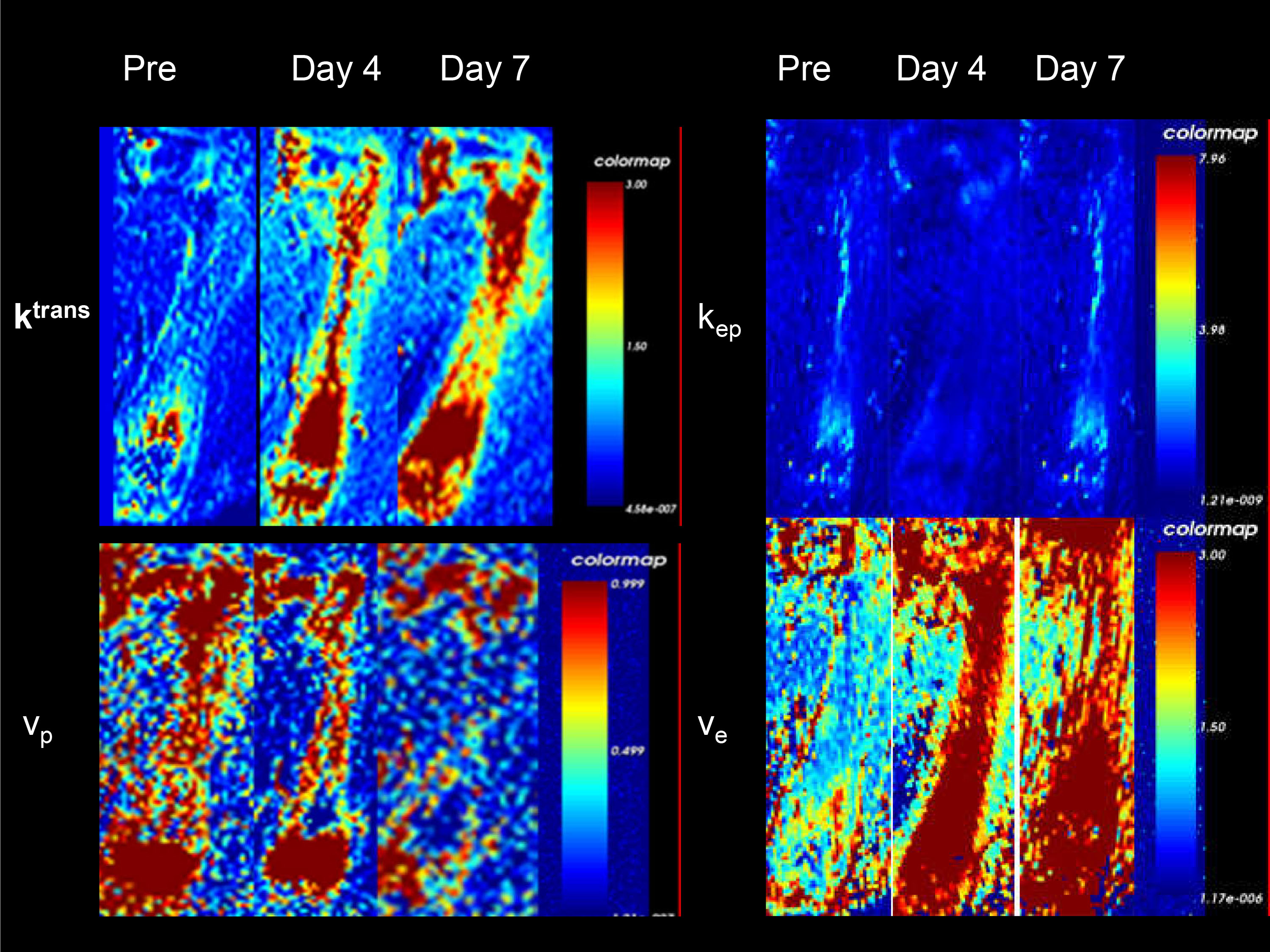
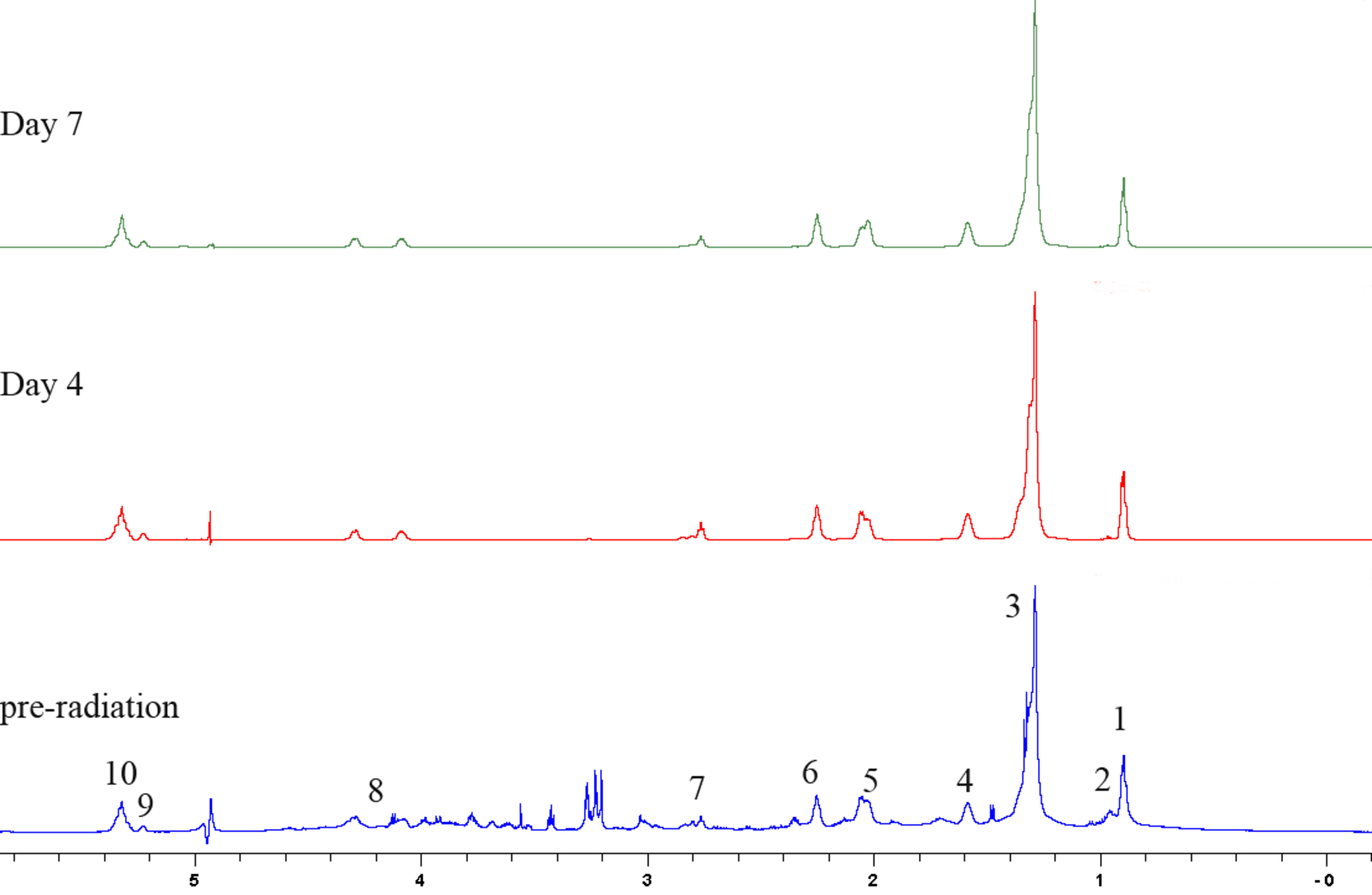
Thirty-six SD rats were randomly divided into irradiation group and normal control group.The irradiation group received 6.0Gy of whole body high-energy x-ray, and permeability imaging using DCE-MRI were performed before irradiation and on days 4 and 7 after irradiation. The following quantitative parameters were measured: the volume transfer constant (Ktrans), the efflux rate constant (kep), blood plasma volume fraction (vp) and extracellular extravascular volume fraction(ve). Rats were then sacrificed and the bone marrow from each rat femur frozen in liquid N2 for high-resolution MAS 1H NMR spectroscopy analysis. Meanwhile, the left rat femur was prepared for determination of bone marrow fat content and microvessel density (MVD) by histopathologic examination.Results
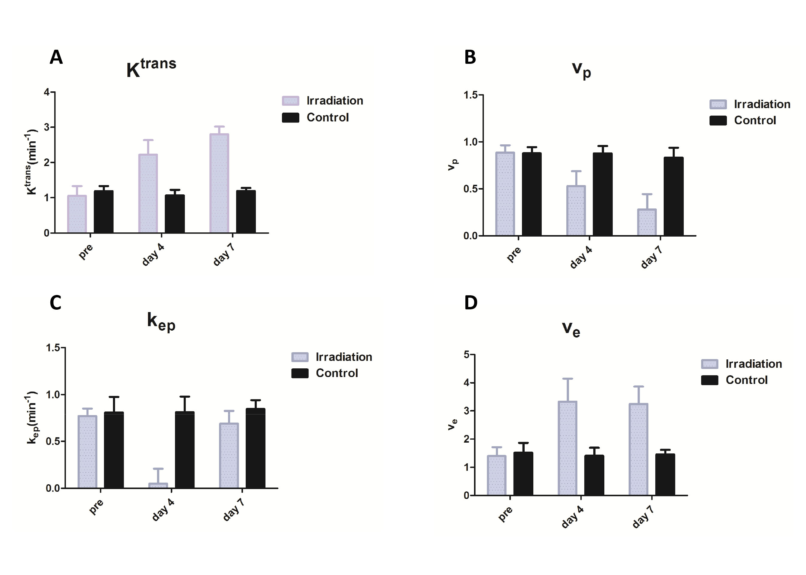
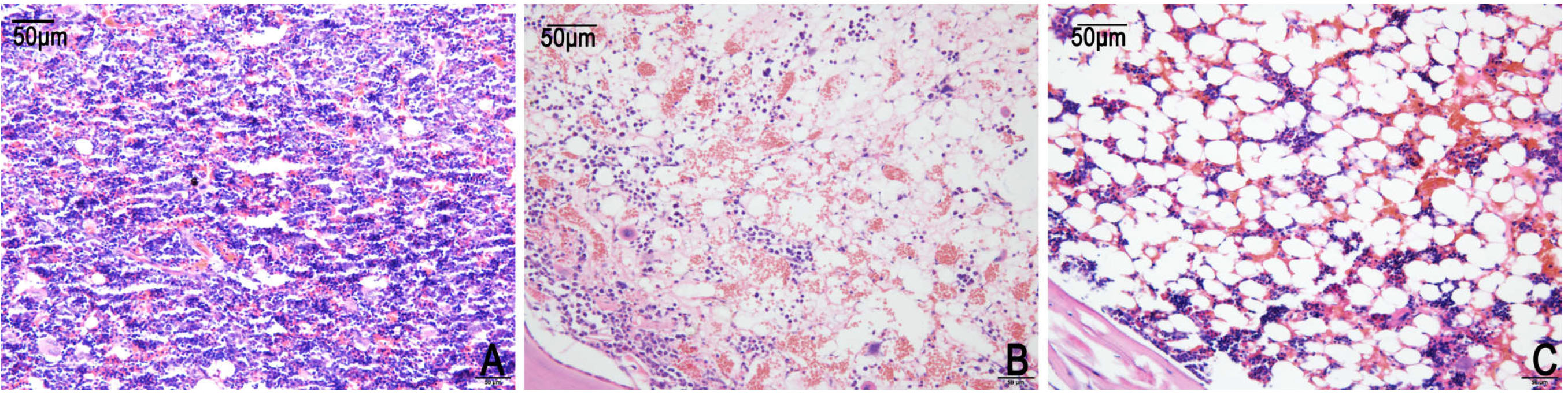
The irradiation group has statistically significant differences in Ktrans, vp, kep and ve values at different time points before and after the irradiation (Fig.1). Fig. 2 shows the Ktrans Values gradually reached a peak on the 7th day after irradiation(A), and vp Values gradually decreased(B). The kep values was reduced to 0.360± 0.049/min (P<0.01) on day 4 and to 0.689± 0.138/min on day 7 (P<0.01) after irradiation (C). the ve Values increased to 3.331± 0.817(P<0.01) on day 4, and remained on day 7 after irradiation (P=0.355) (D). Meanwhile, adipocytes gradually reached a peak on the 7th day after irradiation(Fig. 3). The bone marrow measured fat content by histopathologic examination at different time showed correlation with Ktrans(r= 0.7393, P<0.01) and vp(r=-0.5841, P<0.05). The bone marrow microvessel density (MVD) showed positive correlation with vp(r=0.6451, P<0.05). Bone marrow fatty acid (FA) composition significantly changed at different time points after the irradiation(Fig.4). The proportion of (n−6) PUFA gradually reached a peak on the 7th day, the proportion of (n−3)PUFA gradually decreased and the proportion of saturated FA gradually increased. After the irradiation,Ktrans at different time showed significant negative correlation with(n−3)PUFA(r=-0.6393, P<0.01), significant positive correlation with (n−6) PUFA(r=0.6841, P<0.05).Discussion
The volume transfer constant Ktrans gradually reached a peak on the 7th day after irradiation, whereas the vp Values gradually decreased. Meanwhile, the bone marrow fat content gradually increased, which indicates that adipocytes accumulation result in the oppression of bone marrow sinusoids. This may be an important cause of the decreases of MVD and vp. MVD shows positive correlation with vp, indicating that vp can reflect the average density and is sensitive to perfused vessels.The proportion of (n−6) PUFA promoting vascular inflammation gradually increased, It indicates that the changes of bone marrow fatty acid(FA) composition after the irradiation may be the important cause of the injury of bone marrow microcirculation.Conclusion
Bone marrow microcirculation perfusion and vascular permeability correlate with fat content at early stage after irradiation. There might be a pathophysiology mechanism based on fat- vascular permeability in the injury of bone marrow microcirculation.Acknowledgements
No acknowledgement found.References
1.Cao X et al, Proc Natl Acad Sci U S A, 108(4):1609-1614(2011).
2. J.F. Griffith et al, Radiology, vol. 241, pp. 831-838(2006) .
3. Zhang, Q et al,Radiat Res 172(5): 607-16(2009).
Figures