5104
bSSFP Elliptical Signal Model With GRAPPA Parallel Imaging for Musculoskeletal Applications1Brigham Young University, Provo, UT, United States, 2Electrical Engineering, Brigham Young University, 3Radiology, University of Utah
Synopsis
Balanced steady-state free precession imaging using the elliptical signal model geometrical solution can be combined with a GRAPPA parallel imaging reconstruction that preserves phase information to shorten scan times.
Purpose
The bSSFP pulse sequence is a rapid steady-state sequence that offers high SNR efficiency and excellent contrast in musculoskeletal applications. However, the sequence is particularly sensitive to B0 field inhomogeneity and often suffers from dark banding artifacts. These artifacts may be reduced by taking multiple images where the bands are shifted by phase cycling and combining them in various ways. It has recently been shown that a geometric solution (GS) is more effective at correcting this artifact than the more commonly used complex sum, sum of squares, and max intensity banding removal methods1.
Unfortunately, GS requires at least 4 phase-cycled images whereas the other more commonly used methods often work adequately with only 2. By utilizing parallel imaging methods, we hope to offset the increase in scan time that is required by GS. However, unlike several of the bSSFP techniques, it is critical to preserve accurate phase information for the GS technique. GRAPPA is a commonly known parallel imaging method that preserves phase information2, which is required to implement the GS. Here we show a combination of the GS and GRAPPA with an acceleration of 2, showing that although 4 images are required for GS, it is still a fast and valid choice for bSSFP imaging.
Methods
Phantom Experiment:
A uniform phantom was scanned at 3T on a Siemens TIM Trio system using a 12-channel head matrix. A phase cycled bSSFP sequence was used to acquire both fully sampled and accelerated data. Fully sampled data was acquired with 4 different phase cycles (0°, 90°, 180°, and 270°), TE/TR = 2.12/4.24 ms, flip angle = 70°, voxel size = 2.34x2.34x5 mm, and matrix size 128 x 64 x 24. Accelerated data was acquired using the same parameters and an acceleration factor R of 2 with 24 ACS lines. Total acquisition time for fully sampled data was 26 s while total acquisition time for the GRAPPA accelerated data was only 18 s. Reconstructions were performed offline using MATLAB to obtain raw k-space data and implement the GS for banding removal.
In-Vivo Experiment:
A knee from a healthy volunteer was scanned at 3T on a Siemens TIM Trio system using an 8-channel knee coil. A phase cycle bSSFP sequence was used to acquire both fully sampled and accelerated data. Fully sampled data was acquired with 4 different phase cycles (0°, 90°, 180°, and 270°), TE/TR = 2.77/5.54 ms, flip angle = 30°, voxel size = 0.94x0.94x1 mm, matrix size 256 x 256 x 8. Accelerated data was acquired using the same parameters and an R=2 with 24 ACS lines. Acquisition times were 57 s (fully sampled) and 31 s (GRAPPA). Reconstructions were performed offline using MATLAB to obtain raw k-space data and implement the GS for banding removal.
Results
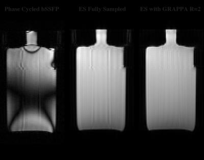
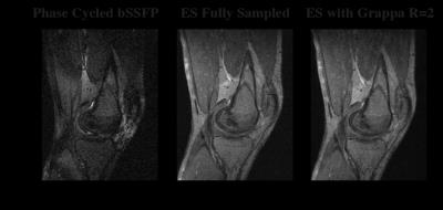
The combination of GRAPPA and GS yielded nearly complete banding removal (Figure 1). Using an acceleration factor of 2, the total acquisition time for the phantom data was 69% that of the fully sampled data. Residual aliasing was also negligible in the GRAPPA accelerated images compared to the fully sampled data. Likewise, the in vivo experiment achieved an acquisition time that was 55% of the fully sampled requirement but had minimal residual aliasing and virtually no banding (Figure 2).Discussion
bSSFP is a sequence often utilized in MSK imaging since it has desirable contrast, quick acquisition times, and high SNR. Traditionally, one of the limitations of this sequence has been residual banding that may limit image utility for diagnosis. Our results demonstrate that these artifacts can be eliminated without a significant increase in acquisition time. Further acceleration could be achieved by increasing the GRAPPA acceleration factor, using 2D GRAPPA for 3D data, or incorporating compressed sensing in conjunction with a parallel imaging technique. For bSSFP images, better removal of aliasing artifacts may also be achieved by using a single GRAPPA kernel over all phase cycles to improve calibration quality3.Acknowledgements
We would like to acknowledge NIH R01EB002524 for providing financial support.References
1. Xiang Q-S, Hoff MN. Banding artifact removal for bSSFP imaging with an elliptical signal model. 2014; MRM 71(3):927-933.
2. Griswold MA et al. Generalized autocalibrating partially parallel acquisitions (GRAPPA). 2002; MRM 47(6):1202-10.
3. Baron C, Jou T, Datta A, Pauly J, Nishimura D. Robust GRAPPA Calibration in Phase Cycled BSSFP. Proc. Intl. Soc. Mag. Reson. Med. 24 (2016):3216.
Figures

