5100
High resolution MRF-FISP with radial acquisition for MSK imaging1School of Electrical & Electronic Engineering, Yonsei University, Seoul, Korea, Republic of
Synopsis
Magnetic resonance fingerprinting (MRF) provides rapid quantification of proton density, T1 and T2 mapping using spiral acquisition. However, for high resolution imaging, spiral imaging can be limiting due to its relatively long readout time. In this study, we implemented high resolution (0.5x0.5mm2) MRF-FISP using golden angle random rotating radial acquisition.
Purpose
Magnetic resonance fingerprinting is a novel technique which provides rapid mapping for proton density, T1 and T2 values1. MRF uses random flip angles and TRs to generate unique signal evolution. Highly undersampled spiral readout acquisition were applied to grant very short scan time. For applications that require high resolution imaging such as MSK, the spiral based approach can be limiting because spiral acquisition requires relatively long readout duration. An alternative approach would be to use a radial acquisition. Studies using radial acquisition in MRF2 have been reported, however, high resolution (e.g. 0.5x0.5mm2) has not been performed. Here, we implemented a high resolution MRF-FISP using golden angle random rotating radial acquisition.Methods
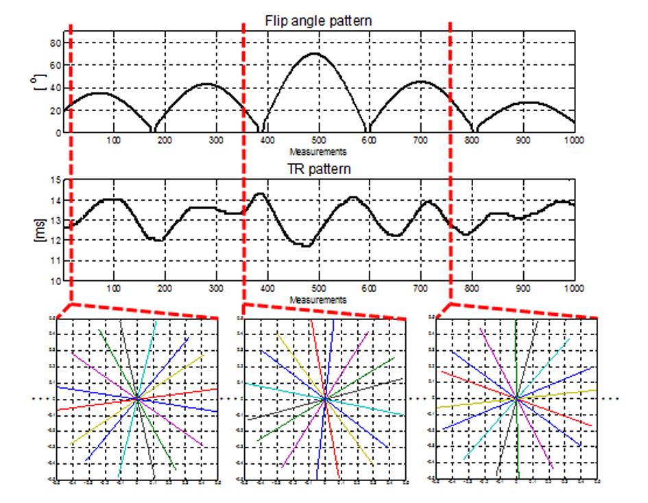
Highly undersampled radial acquisition with Golden angle method3 was applied to the MRF scheme with random rotation at each TR. 4, 8, 31 spokes for each images were acquired for the comparison. The FA and TR patterns shown in Figure 1 were the same as the patterns reported from the original MRF FISP paper4. 3T Siemens scanner (Tim Trio) were used to acquire data with knee coil. Other scan parameters: 0.5x0.5mm2 resolution 512x512 mm2 FOV, 5 mm slice thickness and 391 Hz/Px bandwidth. Spoiler gradients were also applied with the spoiling momentum set to 8π along the slice direction. The scan times were 52 sec/slice, 1 min 45 sec/slice and 6 min 47 sec/slice for each 4, 8, 31 spokes/image acquisition. Each under sampled images were reconstructed using NUFFT5. All experiments were performed on a healthy volunteer (IRB-approved).
A total of 501 isochromats were used to generate MRF-FISP dictionary based on the Bloch simulation. The dictionary had T1 : [100:20:1500] ms, T2 : [3:3:150, 155:5:250] ms, off resonance : [-498:5:-400, -50:5:50] Hz. In the original MRF-FISP paper, the dictionary was generated without off-resonance term because T1 and T2 accuracy were not affected by B0 field inhomogeneity in MRF-FISP. However, in this study, two ranges of off-resonance were added in dictionary to separate water and fat region.
Results
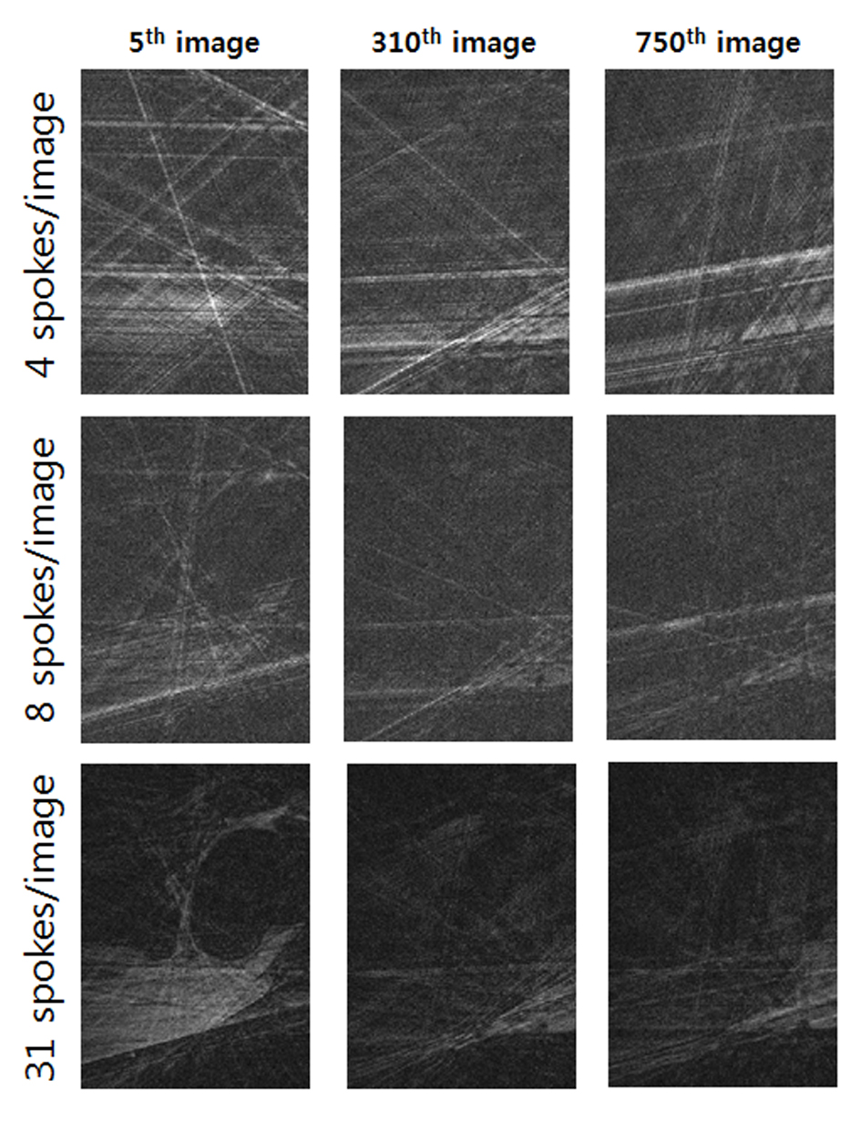
Figure 2 shows several examples of the reconstructed images as different number of spokes/image. The outer region of the result images were masked due to the lack of sensitivity of knee coil. Severe undersampling artifacts were shown in the images reconstructed with only 4 spokes/image.
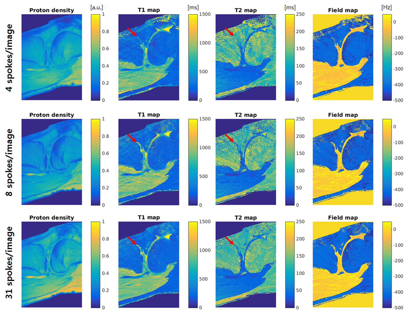
Figure 3 shows the matched results as different number of spokes/image acquisition. Although the proposed method has very high in-plane resolution, the proton density, T1 and T2 maps show clear quantitative results in both 8 and 31 spokes/image acquisition. However, some details were disappeared in the results using 4 spokes/image acquisition.
Smooth and small field variation were not shown in the field map, because MRF-FISP is inherently insensitive to field inhomogeneity. Chemical shift effect of fat was presented as off-resonance value which was resulted in the -498~-400 Hz range in the field map.
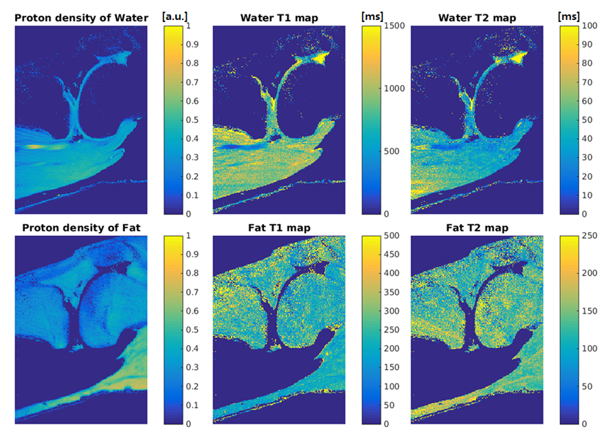
Figure 4 shows the water/fat separation results (8 spokes/image data) based on acquired field map. The quantitative T1 and T2 values of both water and fat were in good agreement with the previous research6.
Discussion
We investigated high resolution MRF-FISP with radial acquisition as different number of spokes/image. However optimization of this parameters need further investigation to improve the result of 4 spokes/image acquisition. Also, partial volume effect due to chemical shift were not dealt with in this study. Further research, such as using OMP8, need to be investigated to overcome this problem.Conclusion
We have presented a rapid high resolution (0.5x0.5mm2) water and fat separated proton density, T1 and T2 mapping using MRF-FISP with random rotating radial acquisition in knee.Acknowledgements
This research was supported by Global PH.D Fellowship Program through the National Research Foundation of Korea(NRF) funded by the Ministry of Education (NRF-2015H1A2A1030698)References
1. Ma D, Gulani V, Seiberlich N, Liu K, Sunshine JL, Duerk JL, Griswold MA. Magnetic resonance fingerprinting. Nature 2013;495:187-192.
2. Cloos MA, Knoll F, Zhao T, Block KT, Bruno M, Wiggins GC, Sodickson DK. Multiparametric imaging with heterogeneous radiofrequency fields. Nat Commun 2016;7:12445.
3. Winkelmann S, Schaeffter T, Koehler T, Eggers H, Doessel O. An optimal radial profile order based on the Golden Ratio for time-resolved MRI. IEEE Trans Med Imaging 2007;26:68-76.
4. Jiang Y, Ma D, Seiberlich N, Gulani V, Griswold MA. MR fingerprinting using fast imaging with steady state precession (FISP) with spiral readout. Magn Reson Med 2015;74:1621-1631.
5. Fessler JA, Sutton BP. Nonuniform fast fourier transforms using minmax interpolation. IEEE Trans Signal Process 2003;51:560–574.
6. Gold GE, Suh B, Sawyer-Glover A, Beaulieu C. Musculoskeletal MRI at 3.0 T: initial clinical experience. AJR Am J Roentgenol 2004;183:1479-1486.
7. Pierre EY, Ma D, Chen Y, Badve C, Griswold MA. Multiscale reconstruction for MR fingerprinting. Magn Reson Med 2016;75:2481-2492.
8. Pati YC, Rezaiifar R, Krishnaprasad P. Orthogonal matching pursuit: Recursive function approximation with applications to wavelet decomposition. 1993. IEEE. p 40-44.
Figures